4.1.1 POJA-L4168+4167+4169+4170+4172+4172+4173
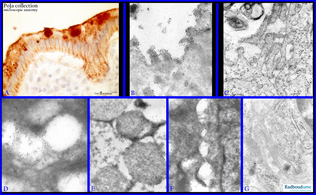
Title: Ultrastructural immune-gold localization of CO-TL1 antibody-defined epitopes in colon epithelium (human)
Description: Stain: (A) CO-TL1 monoclonal antibody immunoperoxidase staining with diaminobenzidin reaction (DAB) and hematoxylin counterstaining. (B-G) Ultracryomicroscopy and Lowicryl-embedding procedure using gold labeled monoclonal antibody CO-TL1.
In (A) it is shown that the CO-TL1-defined colon-specific antigen is localized in all colorectal epithelial cells, with strongest expression in the goblet cells.
(B-G): shows the ultrastructural localization of the antigen with 10 nm gold particles.
(B): Positive label on microvilli of colonocytes as well as in the most apical small secretion granules.
(C): Lowicryl-embedded specimen of colon tissue showing a precise localization of colon-specific antigen on the interdigitating cell membranes.
(D): Positive gold label staining of the large Golgi vesicles.
(E): Immunogold labeling of the secretion granules in goblet cells.
(F): Dense localization of the immunogold label on the interdigitating lateral membranes of two adjacent colonocytes.
(G): Discrete localization of the gold label in the Golgi vesicles (Lowicryl embedding).
Keywords/Mesh: colon, CO-TL1, colon-specific antigen, electron microscopy, immuno gold labeling, histology, POJA collection

