
2.4 POJA-L1090. Colon (‘gut-associated lymphatic tissue’ or GALT) (human)
2.4 POJA-L1090
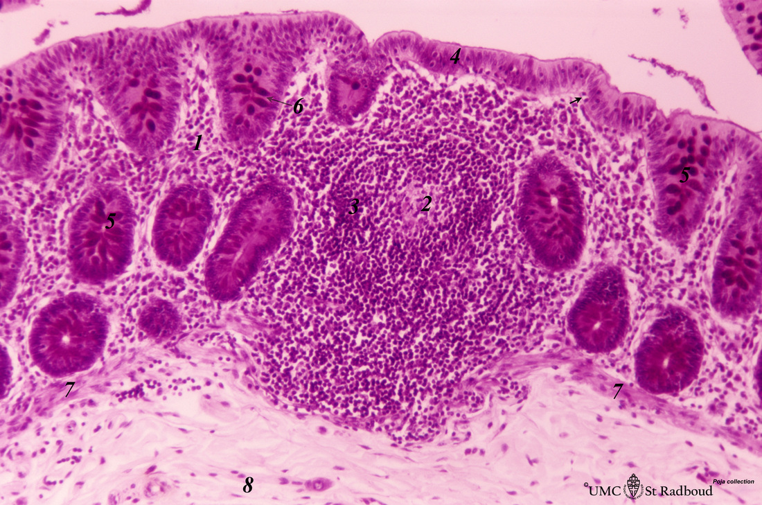
Title: Colon (‘gut-associated lymphatic tissue’ or GALT) (human)
Description: Stain: Hematoxylin-PAS. Solitary lymphatic nodule in colon (see also Digestive System: Colon).
A large amount of non-encapsulated diffuse lymphatic tissue or mucosa-associated lymphatic tissue (MALT) is located in the subepithelial lamina propria of the colon and called gut-associated lymphatic tissue (GALT).
A secondary lymphatic nodule in the colon extends throughout the proper lamina (1). The nodule is similar to that found in a lymph node with germinal centre (2) and darker-stained mantle zone (3). The lumen is lined by a layer of columnar epithelial cells (4) with diapedesis of lymphocytes (→).
(5) Short crypts with columnar absorptive epithelial cells and interspersed bluish red-stained goblet cells (→, 6). A thin layer of lamina muscularis mucosae is visible (7). (8) Submucosa layer composed as loose connective tissue.
Keywords/Mesh: lymphatic tissue, MALT, GALT, colon, lymphoepithelial tissue, reticular tissue, histology, POJA collection