7.3 POJA-L1707+1706+1705
Title: Complete hydatidiform mole (human)
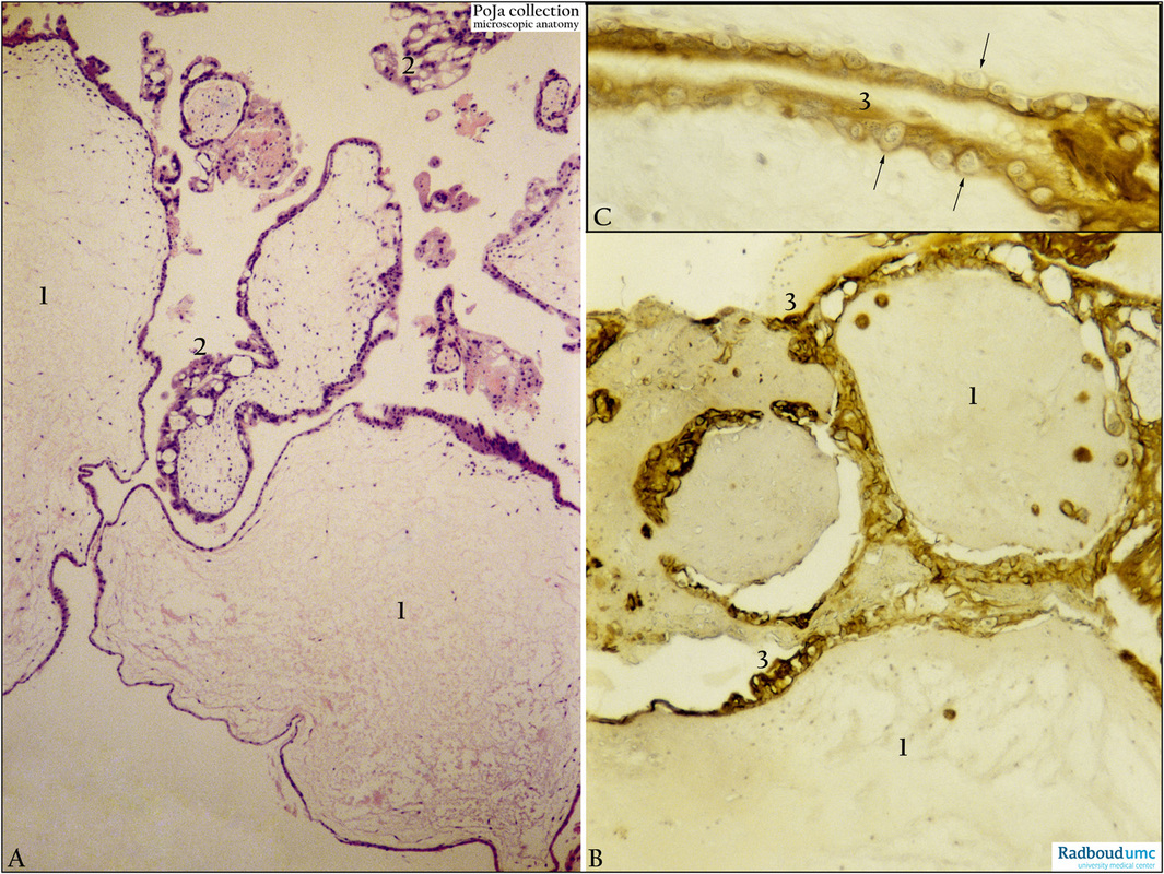
Description: Stain: (A) Hematoxylin-eosin. (B+C) Immunoperoxidase staining with diaminobenzidin (DAB) and hematoxylin counterstaining for keratin 7 (OVTL 12-30 antibody).
(A)Trophoblast cells surround the dilated chorionic villi (1), with centrally large amounts of edematous mesenchymal stroma with almost no blood vessels. The villus lining shows irregular proliferation resulting in large solid cell masses (2). (Partly by courtesy of G. P. Vooijs MD PhD, former Head of the Department of Pathology, Radboud university medical center, Nijmegen, The Netherlands).
(B+C) Keratin 7-staining is distinct for lining syncytiotrophoblast cells (3) and strongly positive for proliferating ones while lining cytotrophoblast cells appear negatively (arrows).
Keywords/Mesh: complete hydatidiform mole, placenta, villus, trophoblast, immunohistochemistry, keratin-7, hydatidiform mole, histology, POJA collection.

