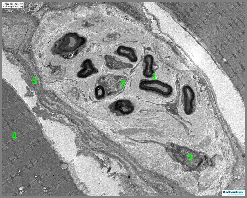
14.1 POJA-L6250 Electron micrograph of myelinated nerve bundle in the perimysium (human)
14.1 POJA-L6250 Electron micrograph of myelinated nerve bundle in the perimysium (human)
(By courtesy of L. Eshuis BSc Section Electron microscopy, Department of Pathology, Radboud university medical center, Nijmegen, The Netherlands)
Title: Electron micrograph of myelinated nerve bundle in the perimysium (human)
Description:
Encapsulated within the perimysium (3) a bundle of myelinated nerve fibres (1) surrounded by the Schwan cell (2), positioned between muscle fibres (4). Note a solitary phagocytic cell (5) with slender projections occurs within the enclosing flattened perineural cells that surround the nerve fibers.
Keywords/Mesh: locomotor system, skeletal muscle, striated muscle, perimysium, interstitium, innervation, electron microscopy, POJA collection

