
2.4 POJA-L1101. Scheme of ileum with Peyer’s patches (‘gut-associated lymphatic tissue or GALT) (dog)
2.4 POJA-L1101
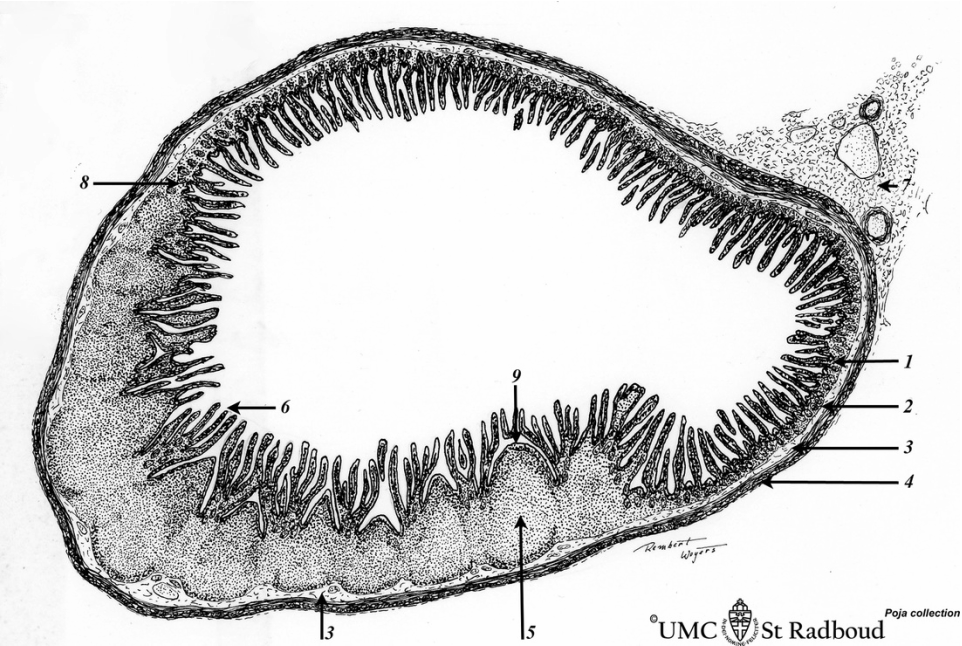
Title: Scheme of ileum with Peyer’s patches (‘gut-associated lymphatic tissue or GALT) (dog)
Description: Survey ileum (see also Digestive System: Ileum)
A large amount of non-encapsulated diffuse lymphatic tissue or mucosa-associated lymphatic tissue (MALT) is located in the subepithelial lamina propria (e.g. respiratory passages, genitourinary tract).
The gut-associated lymphatic tissues (GALT) are found in the oesophagus, small intestine and in the large intestine. In the ileum so-called Peyer’patches are large aggregations of MALT (about 200-400 nodules) extending from the proper lamina into the submucosa, often bulging (domes) into the lumen of the gut. The nodules are similar to those found in the lymph node with darker-stained caps oriented towards the epithelium of the gut. Dome areas (9) bulge from the lymphatic tissue of the submucosa into the intestinal lumen. Here, the epithelium of the dome areas does not display microvilli or crypts or mucous-producing goblet cells. Instead of microvilli these specialized epithelial cells show microfolds at their surfaces (microfold cells or M-cells). These M-cells function as antigen-transporter cells towards the underlying follicles.
Keywords/Mesh: lymphatic tissue, MALT, GALT, ileum, Peyer’s patch, lymphoepithelial tissue, reticular tissue, histology, POJA collection
Title: Scheme of ileum with Peyer’s patches (‘gut-associated lymphatic tissue or GALT) (dog)
Description: Survey ileum (see also Digestive System: Ileum)
A large amount of non-encapsulated diffuse lymphatic tissue or mucosa-associated lymphatic tissue (MALT) is located in the subepithelial lamina propria (e.g. respiratory passages, genitourinary tract).
The gut-associated lymphatic tissues (GALT) are found in the oesophagus, small intestine and in the large intestine. In the ileum so-called Peyer’patches are large aggregations of MALT (about 200-400 nodules) extending from the proper lamina into the submucosa, often bulging (domes) into the lumen of the gut. The nodules are similar to those found in the lymph node with darker-stained caps oriented towards the epithelium of the gut. Dome areas (9) bulge from the lymphatic tissue of the submucosa into the intestinal lumen. Here, the epithelium of the dome areas does not display microvilli or crypts or mucous-producing goblet cells. Instead of microvilli these specialized epithelial cells show microfolds at their surfaces (microfold cells or M-cells). These M-cells function as antigen-transporter cells towards the underlying follicles.
- mucosal layer with proper lamina
- muscular layer
- submucosal layer
- tunica muscularis
- secondary lymphatic nodules with germinal centres in proper lamina as well as in submucosa
- villus
- mesentery
- crypt
- dome
Keywords/Mesh: lymphatic tissue, MALT, GALT, ileum, Peyer’s patch, lymphoepithelial tissue, reticular tissue, histology, POJA collection