
13.1 POJA-L4611+4612+4631+La-0317/4615+4616 Transitional arteries (human)
13.1 POJA-L4611+4612+4631+La-0317/4615+4616
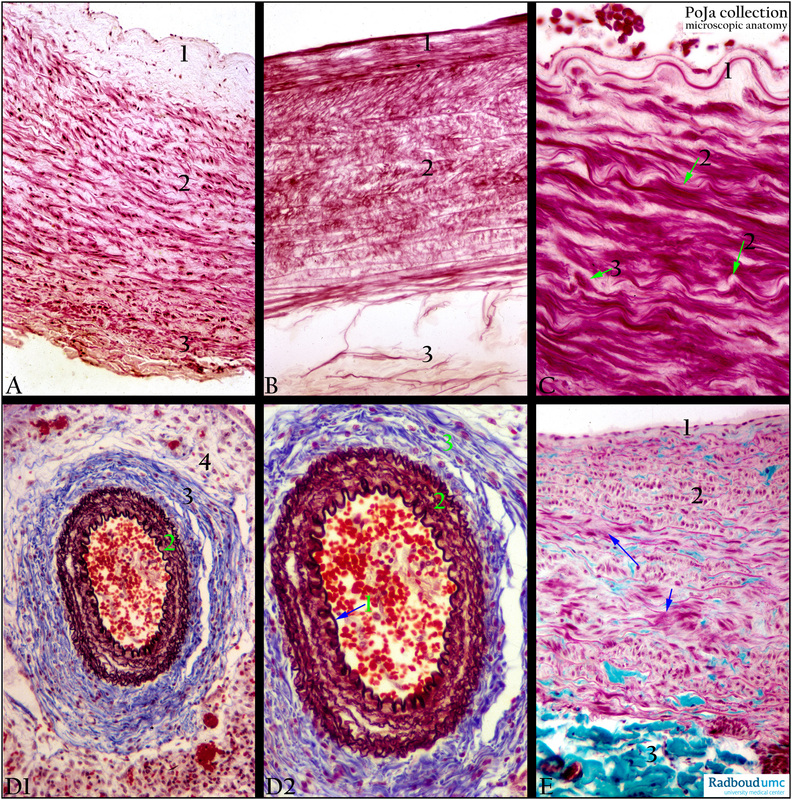
Title: Transitional arteries (human)
Description:
(A, B, C): Staining: (A) Haematoxylin-eosin, (B) Orcein, (C) Azan. Axillar arteries are transitional forms (hybrid arteries) of elastic arteries into muscular types. The elastic membranes in these arteries are thinner and shorter but their number is increased. The SMC (smooth muscle cell) layer of the media is less developed. The intima shows longitudinal oriented SMCs. Generally, arteries exposed to multiple bending contain relatively many longitudinal SMCs (e.g. popliteal artery, axillar artery).
(A-B): (1) Intima (widened due to some edema and ageing). (2) Media with elastic lamellae as well as SMCs. (3) Adventitia.
(C): Proximal part of axillar artery, detail from intima and upper part of media. (1) Internal elastic membrane (IEL). (2) Elastic lamellae in media. (3) SMC in media.
(D1-D2-E): (D) Mallory, neonate. (E) Trichrome (Goldner), adult. (D1-D2): Survey and detail (1) Intima with internal elastic lamina (IEL). (2) Elastic membranes in media. (3) Adventitia. (4) Adventitia continuous with surrounding connective tissue.
(E): Cross-section of vessel: (1) Intima, (2) Elastic membranes alternate with layers of SMCs (arrow) in media. Variation of circular and cross-sectioned SMCs in the wall is the result of the twisted blood vessel. (3) Adventitia.
Background:
Pulmonary arteries: Due to reduction of muscular and elastic elements the extrapulmonary artery (1mm diameter) is an elastic artery with a thinner wall. Intralobularly the artery becomes muscular. The blood pressure is low, with blood low in oxygen and high in carbon dioxide. Apart from elastic membranes and networks of elastic fibres there is an increased amount of collagen, while the SMCs are less regular orientated. Proximally the pulmonary artery contains cardiomyocytes in its wall. On the other hand the pulmonary vein contains oxygen-rich blood and has a well-developed SMC media. In the mouse cardiomyocytes are also found in the media.
Keywords/Mesh: cardiovascular system, vascularisation, blood vessel, transitional artery, hybrid artery, elastic artery , muscular artery, pulmonary artery, histology, POJA collection