
1.1 POJA-L838. Erythroblastic island (bone marrow, rabbit)
1.1 POJA-L838
Title: Erythroblastic island (bone marrow, rabbit)
Description: Electron microscopy.
In the bone marrow close associations of developing red blood cells with reticular cells are required during erythropoiesis. Different stages of erythroblasts (2, 3) are exposed in close vicinity of a reticular cell (1).
(4): Reticulocyte.
The centrally localized phagocytic reticular cell (1) has many long cytoplasmic extensions that form networks with identical cells within the bone marrow. The cell has a characteristic electron-light nucleus. The cytoplasm contains moderate amount of organelles however with a distinct variety of lysosomal structures and shows upload with phagocytised remnants of expulsed erythroblastic nuclei and other degraded material (arrows, ↓).
Keywords/Mesh: blood, bone marrow, erythropoiesis, reticular cell, erythroblast, reticulocyte, histology, electron microscopy, POJA collection
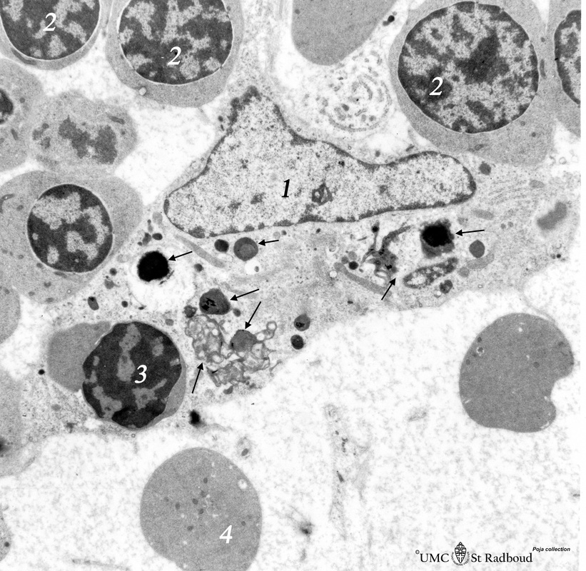