
2.3 POJA-L1033. Lymph node (human)
2.3 POJA-L1033
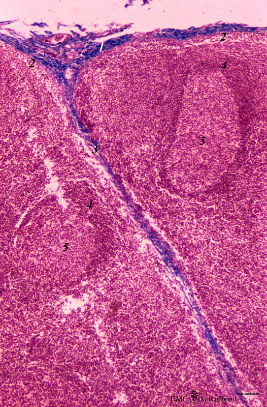
Title: Lymph node (human)
Description: Stain: Azan.
Part of the cortex of a lymph node with the capsule (1) and subcapsular (or marginal) sinus (2) filled with lymphocytes. From the blue-stained dense capsule a long trabecula flanked by paratrabecular (intermediate) sinuses penetrates between the paracortical areas (3). Paratrabecular sinuses arise from the subcapsular sinus and continue into the medulla as branching medullary sinuses surrounded by medullary cords.
Below the subcapsular sinus the crescent or corona (4) of a superficial secondary follicle with a germinal centre (5), at the left a deeper localized secondary follicle. The follicles contain predominantly B lymphocytes, the paracortex between and below the follicles mostly contain T cells. Each area has its own type of antigen-presenting cells or (inter)digitating cells (1) DC’s.
Keywords/Mesh: lymphatic tissue, lymph node, lymphatic follicle, paracortex, reticular tissue, histology, POJA collection