
4.1.1 POJA-L3638+3959+3971+3977. Fundus area of the stomach (human, dog, cat)
4.1.1 POJA-L-3638+3959+3971+3977
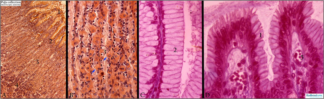
Title: Fundus area of the stomach (human, dog, cat)
Description: Stain: (A, B). Alum hematoxylin-eosin mucocarmin (cat). (C, D) Azan, human, dog).
The survey of the fundus of the stomach in (A) shows the infoldings of the gastric surface epithelium (1) into foveolae (2) that subsequently extent into long slender tubules of the gastric glands (3). The transition area from foveolae (2) to the tubules comprises the so called neck cells (C) that produce mucus. They are tall columnar cells.
The middle zone of the gland (3) comprises intensely stained parietal cells (arrow, ↘) that secrete HCl (and intrinsic factor) for which numerous mitochondria are required. The mitochondria are responsible for the intense histological staining of those cells.
The bottom of the gland tubules are stained darkly (4) and contain largely the chief cells that generate the proteolytic enzymes such as pepsinogen and lipases. Spread throughout, but not shown here, are the enteroendocrine cells producing hormones as glucagon and gastrin.
Note the small capillaries (5) in the top of the foveolae.
Keywords/Mesh: stomach, fundus, parietal cells, chief cells, histology, POJA collection