6.3 POJA-L3811+2739
Title: Seminal vesicle
Description:
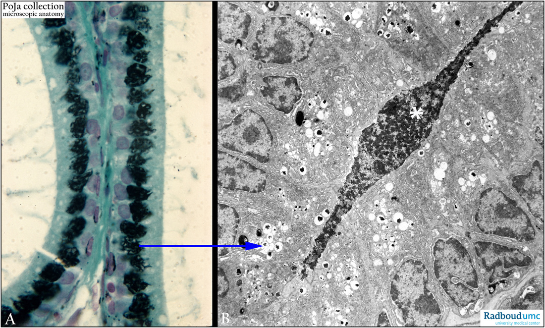
(A) Osmium impregnation, rat. (B) Electron microscopy, gerbil.
Detail of the epithelial cells in two cross-sections of the seminal vesicle (or glandula vesiculosa).
The osmium impregnation in (A) highlights the darkly stained cisterns and vesicles of the Golgi factory
detailed in the electron micrograph (B). Note the supranuclear Golgi areas and secretory vesicles in
the epithelial cells in (B). The lumen contain heterogeneous dense-stained secretory material (*), containing minerals.
Keywords/Mesh: seminal vesicle, seminal gland, glandula vesiculosa, histology, electron microscopy, POJA collection

