
4.2.1 POJA-L3737
Latex uptake by liver Kupffer cell (mouse)
4.2.1 POJA-L3737
Title: Latex uptake by liver Kupffer cell (mouse)
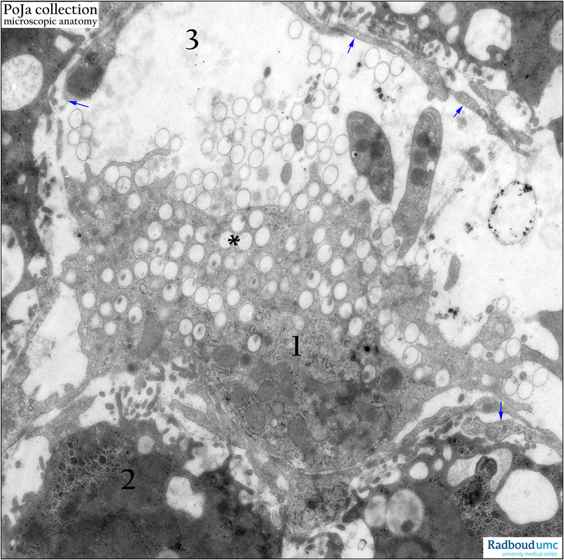
Description: Electron micrograph of a Kupffer cell (1) that has ingested perfused latex particles (*) after a few minutes. The latex particles are electron-lucent. The cell is localized in the sinusoid space (3) and borders the adjacent liver cells (2). Arrows (↘) indicate the long slender processes of the Kupffer cell. Note two thrombocytes.
Keywords/Mesh: liver cell, Kupffer cell, sinusoid, latex particles, phagocytosis, electron microscopy, POJA collection
Title: Latex uptake by liver Kupffer cell (mouse)
Description: Electron micrograph of a Kupffer cell (1) that has ingested perfused latex particles (*) after a few minutes. The latex particles are electron-lucent. The cell is localized in the sinusoid space (3) and borders the adjacent liver cells (2). Arrows (↘) indicate the long slender processes of the Kupffer cell. Note two thrombocytes.
Keywords/Mesh: liver cell, Kupffer cell, sinusoid, latex particles, phagocytosis, electron microscopy, POJA collection