
12.1.5 POJA-L4407+2960+
3889+2592.
Eye lens
12.1.5 POJA-L4407+2960+3889+2592
Title: Eye lens
Description:
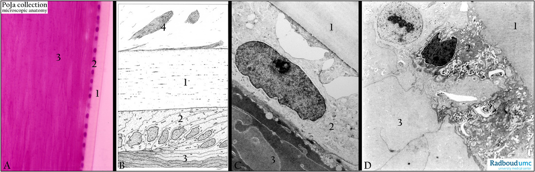
(A): Lens, stain hematoxylin-eosin, human. (1) Capsule being a thick amorph basement membrane consists of collagen IV and glycosaminoglycans. (2) Anterior lens epithelium. (3) Lens fibers.
(B): Electron microscopy scheme of the lens equator zone, human. (1) Thick capsule. At the equator the cuboidal cells (2)
elongate but still contain their nuclei. (3) Towards the central (=nucleus) part of the lens the elongated lens cells (=lens fibers)
lose their organelles and become fine granulated. (4) Zonular fibers from the ciliary body merge with the capsule.
(C, D): Electron micrograph of the lens capsule and anterior epithelium, human (C) respectively bovine (D).
Identical numbers and structures are observed in (C, D). Note similarity of thick fine-granular basal lamina or the capsule (1)
in bovine and human. (2) Anterior epithelial cells, the electron-light cells are the vital ones the darker ones are less functional
or in the process of apoptosis. (3) Characteristic aspect of tightly connected anucleated lens fibers, the granular cytoplasm
hardly contains organelles.
In (B) the very first layer of lens fibers close to the epithelial cells show electron-lighter vesicles in conjunction with their cell
membranes. This phenomenon is commonly observed in eyes of elderly people.
Keywords/Mesh: eye, lens, lens fiber, capsule, histology, electron microscopy, POJA collection
Title: Eye lens
Description:
(A): Lens, stain hematoxylin-eosin, human. (1) Capsule being a thick amorph basement membrane consists of collagen IV and glycosaminoglycans. (2) Anterior lens epithelium. (3) Lens fibers.
(B): Electron microscopy scheme of the lens equator zone, human. (1) Thick capsule. At the equator the cuboidal cells (2)
elongate but still contain their nuclei. (3) Towards the central (=nucleus) part of the lens the elongated lens cells (=lens fibers)
lose their organelles and become fine granulated. (4) Zonular fibers from the ciliary body merge with the capsule.
(C, D): Electron micrograph of the lens capsule and anterior epithelium, human (C) respectively bovine (D).
Identical numbers and structures are observed in (C, D). Note similarity of thick fine-granular basal lamina or the capsule (1)
in bovine and human. (2) Anterior epithelial cells, the electron-light cells are the vital ones the darker ones are less functional
or in the process of apoptosis. (3) Characteristic aspect of tightly connected anucleated lens fibers, the granular cytoplasm
hardly contains organelles.
In (B) the very first layer of lens fibers close to the epithelial cells show electron-lighter vesicles in conjunction with their cell
membranes. This phenomenon is commonly observed in eyes of elderly people.
Keywords/Mesh: eye, lens, lens fiber, capsule, histology, electron microscopy, POJA collection