13.1 POJA-L4677+4678+4679+3636
Title: Mid-sized lymphatic vessels (human)
Description:
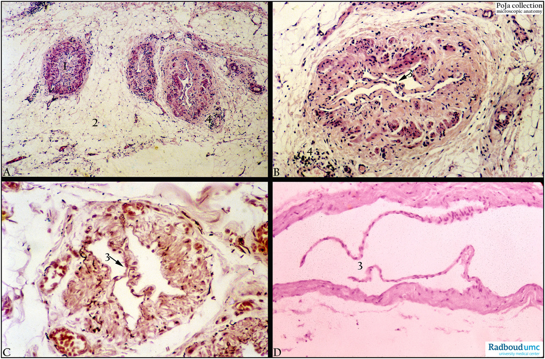
(A, B, C): Lymphatic vessels close to a lymph node in mesentery, (A, B, D) Haematoxylin-eosin stained and (C) orcein-stained. At low magnification (A) three large lymphatic vessels (1) embedded in adipose tissue (2). Note the densely stained irregular bundles of smooth muscle cells (SMC) in the wall of the lymphatic vessels. Detail shows distinct valves (3, arrows). (4) Lymphoid infiltration. Orcein stain reveals irregularly distributed elastic fibres, densely stained in these lymphatic vessels.
(D): Mid-sized lymphatic vessel with long valves (3) (lymph node region), Haematoxylin-eosin. Generally, walls of lymphatic vessels are thin and valves are commonly present. Valves are connective tissue foldings covered with the endothelial lining. Larger lymphatic vessels might show a tripartite wall (e.g. thoracic duct).
Background: Lymphatic vessels (or collecting lymphatics) include the tubular lymphatic capillaries followed by larger collecting vessels and subsequently the right lymphatic duct and the left lymphatic duct (= thoracic duct). The walls of lymphatic vessels are similar to those of small veins and consist of a thin intimal layer, middle layer (concentric-like arranged smooth muscle layers) and fibroelastic adventitial layer. There are abundant valves in the lymphatic vessels, they support the lymph flow without reflux in one direction. The valves use specialised junctions together with anchoring filaments to allow an unidirectional flow to the primary vessels. The collecting vessels propel the lymph by combined actions of valves and SMC in the vessel wall. The capillaries are blind-ended and the valves at the ends of capillaries use specialised junctions together with anchoring filaments to allow a unidirectional flow to the primary vessels. Two valve systems are used to achieve this one directional flow, a primary and a secondary valve system (Bull Math Biol. 2013 Jan; 75(1):49-81. doi: 10.1007/s11538-012-9793-2. Epub 2012 Nov 17. A model for fluid drainage by the lymphatic system. Heppell C1, Richardson G, Roose T. http://www.ncbi.nlm.nih.gov/pubmed/23161129 )
Further readings on Glymphatic system: see: POJA-L4750
Keywords/Mesh: cardiovascular system, vascularisation, lymphatic system, mesentery, lymphatic vessel, valve, lymph, histology, POJA collection

