
6.2 POJA-L2713+La0199.
Efferent ductule and epididymal duct
6.2 POJA-L2713+La0199
Title: Efferent ductule and epididymal duct
Description:
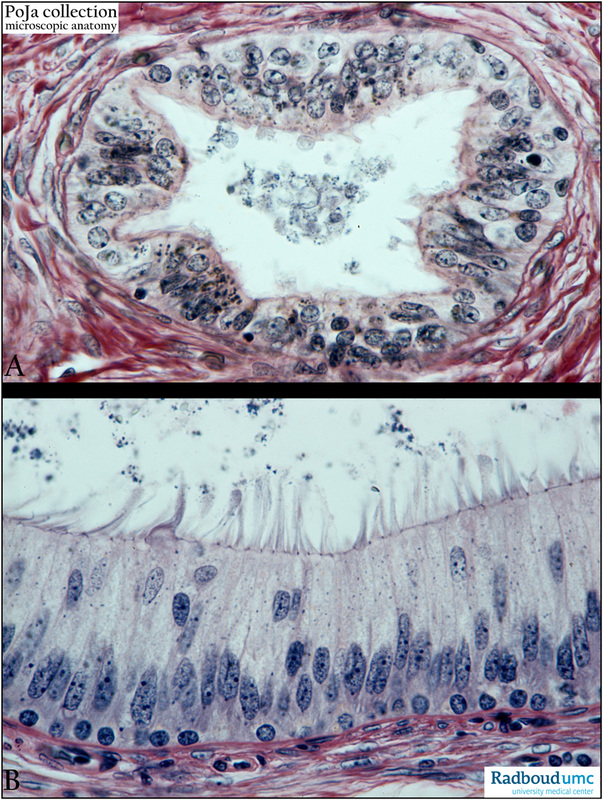
(A) Efferent ductule, human. (B) Epididymal duct, stain Weigert hematoxylin, human.
The efferent ductule (A) shows a pseudostratified epithelial cell lining with irregular protrusions that bear tufts of stereocilia or kinocilia/short microvilli. The efferent ductules continue into the epididymal duct (B).
The epididymal duct is lined with a two-layered pseudostratified epithelium and bears stereocilia sticking together in tufts (B).
The two layers comprise a basal cell layer with small, round cells replacing the layer of columnar principal cells with oval nuclei.
Both the tubule and duct are surrounded with smooth muscle cells/myofibroblasts.
In the lumina intact as well as remnants of spermatozoa are found.
Keywords/Mesh: testis, epididymis, efferent ductule, epididymal duct, histology, POJA collection
Title: Efferent ductule and epididymal duct
Description:
(A) Efferent ductule, human. (B) Epididymal duct, stain Weigert hematoxylin, human.
The efferent ductule (A) shows a pseudostratified epithelial cell lining with irregular protrusions that bear tufts of stereocilia or kinocilia/short microvilli. The efferent ductules continue into the epididymal duct (B).
The epididymal duct is lined with a two-layered pseudostratified epithelium and bears stereocilia sticking together in tufts (B).
The two layers comprise a basal cell layer with small, round cells replacing the layer of columnar principal cells with oval nuclei.
Both the tubule and duct are surrounded with smooth muscle cells/myofibroblasts.
In the lumina intact as well as remnants of spermatozoa are found.
Keywords/Mesh: testis, epididymis, efferent ductule, epididymal duct, histology, POJA collection