POJA-L1289+1297
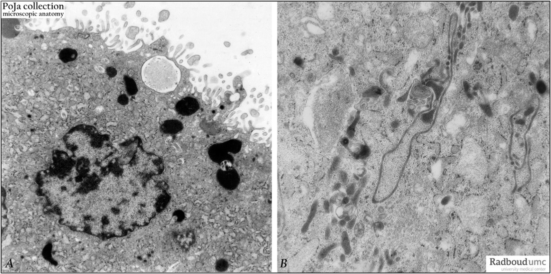
Title: Electron microscopy of syncytiotrophoblast cells in tertiary villus (human placenta, midpregnancy)
Description:
The left photograph (A) shows the apical cytoplasm of a syncytiotrophoblast cell (STC, nucleus). The free cell surface displays small protrusions and a characteristic pattern of differently shaped microvilli; pinocytotic invaginations and a single macropinocytotic vacuole. Note the apical localized electron-dense lysosomal structures also visible at the light-microscopical level. The cytoplasm is stuffed with endoplasmic reticulum and all types of organelles.
In the right picture (B) electron-grey lysosomal structures as well as peculiar long, thin loops of endocytotic structures as well as interconnecting clusters of lysosomal and endocytotic elements, illustrating high phagocytic ability of these STCs.
Keywords/Mesh: placenta , tertiary villi , syncytiotrophoblast, electron microscopy, chorionic villi, trophoblast, histology, POJA collection.

