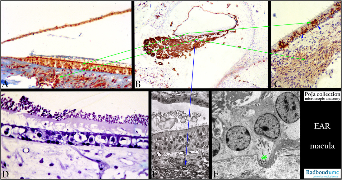
12.2.4.2 POJA-L3424+3409+2638+3389+La0122+3444
Title: Maculae of saccule and utricle in the inner ear
Description:
(A): Macula sacculi, stain Azan, guinea pig. The macula is situated vertically on the wall of the saccule and is attached to the periost of the bony labyrinth. Within the sensory epithelium nuclei of supporing cells remain at the basal side while nuclei of hair cells localise more at the top. The apical borders appear denser-stained due to the cuticular plates of the hair cells with the stubbed stereocilia close to remnants of the otolithic membrane. Below the epithelium thick bundles of myelinated nerve fibres.
(B): Survey of macula, immunoperoxidase staining with AEC and antibodies against collagen IV, 4 days postnatal rat. Note the collagen IV containing basement membranes embedded nerve fibre bundle. The basement membrane of the membranous vestibular labyrinth is also strongly positive, as well as the scattered blood vessels. The cartilage labyrinth represents the future bony labyrinth.
(C): Macula utriculi, immunoperoxidase staining with AEC and antibodies against neurofilament, nervus utriculus, 10 days postnatal rat. Both the nerve fibres and the branched unmyelinated nerve endings between the epithelial sensory hair cells are stained positively.
Striking are the type I hair cells (blue arrowheads) enclosed by the distinctly positive stained basket-like wrapping of the afferent
dendritic projections (zoom in).
(D): Macula sacculi, stain toluidine blue semi-thin plastic section, rat. Note the “stones” (otoconia, statoconia; in fish is called otoliths) resting on a stiff mucous jelly dropped on top of the sensory hair cells.
(E): Macula sacculi, stain toluidine blue semi-thin plastic section (phase contrast black–white), rat. A macula consists of a small sensory
receptor area with type I/type II hair cells and supporting cells covered by a gelatinous glycoprotein layer. Each hair cell has about 40-70 cilia. The stereociliary tips are normally embedded in the so-called otolithic membrane, a jelly-like otoconia-containing substance that contains glycoproteins and a.o. alpha- and beta-tectorin proteins. Due to preparation procedures retraction of the acellular membrane from the stereociliary tips is shown. This image illustrates the crystals (otoconia) or ear dust resting on the otolithic membrane on top of the multilayered epithelium that comprises both sensory cells and supportive cells.
Otoconia contain an inner core made of otoconins (glycoproteins) and proteoglycans while the outer surface consists mostly of precipitated CaCO3. They add to the density of the membrane contributing in sensing motion and gravity.
Unmyelinated nerve terminals within the sensory epithelium continue below the basement membrane as myelinated (densely stained) nerve fibres.
Note at the opposite site of the macula non-sensory epithelial cells lining the membranous duct.
(F): Macula sacculi, electron micrograph, rat. Nerve ending (*) upon entering the neuroepithelium of the macula loses its myelin.
Keywords/Mesh: inner ear, macula, saccule, utricle, otoconium, statoconium, collagen IV, neurofilament, histology, electron microscopy, POJA collection

