4.2.1 POJA-L3786+3781+3783
Title: Mallory bodies as examples of cellular pathology of the liver (human)
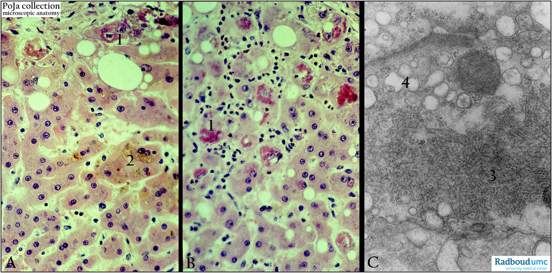
Description: (A, B) stain Hematoxylin-eosin. (C) Electron micrograph.
The intracytoplasmic Mallory bodies are stored cytokeratin aggregates (A, 1; B, 1 and C, 3) modified to advanced glycation end products (AGEs).
(A, 2) Pigment formation. The Mallory bodies occur in several hepatic diseases such as viral hepatitis and alcoholic hepatitis. Note the large amount of neutrophils infiltrated in the area with Mallory bodies (satellitosis).
Keywords/Mesh: liver cell, Mallory bodies, cytokeratin, electron microscopy, histology, POJA collection

