2.2 POJA-L1081+1054
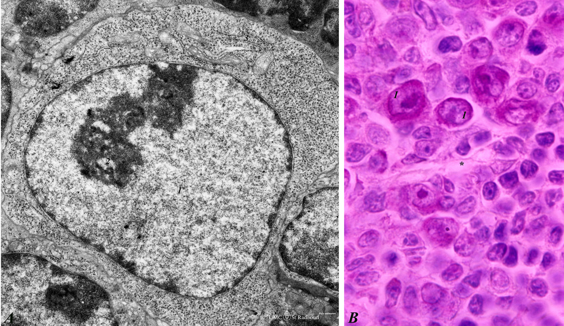
Title: Lymphoblast in splenic white pulp (rat, human)
Description: Electron microscopy (A, rat) and Methyl Green (B, human).
Upon antigenic stimulation the lymphocytes in the germinal centre proliferate and generate activated B cells or lymphoblasts which seed towards marginal zone and red pulp while differentiating. Due to the increased number of lymphoblasts, reticular cells and macrophages the germinal centre of secondary nodules appears lighter-stained. This lymphoblast contains a large nucleus (1) and a distinct nucleolus (2) as well as the numerous polysomes. (3) Area of few endoplasmic profiles and some mitochondria. The lymphoblasts in B are stained red due to their pyroninophylia because of the extended RER and polyribosomes. (*) indicates the marginal zone sinusoids separating the germinal centre area above from the marginal zone and red pulp below.
Keywords/Mesh: lymphatic tissue, spleen, lymphoblast, histology, electron microscopy, POJA collection

