
16.1.3 POJA-L7109+7108+7101+7097 Endochondral ossification in foetus 4
16.1.3 POJA-L7109+7108+7101+7097 Endochondral ossification in foetus 4
16.1.3 POJA-L7109+7108+7101+7097 Endochondral ossification in foetus 4
Title: Endochondral ossification in foetus 4
Description:
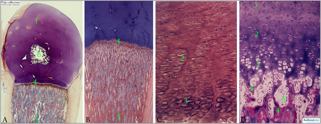
(A-D): Long bone, foetal, human, haematoxylin-eosin, Azan staining.
(1): Epiphysis.
(2): Zone of proliferation.
(3): Zone of hypertrophy.
(4): Ossification zone.
(5): Bone marrow.
(6): Secondary ossification centre.
The epiphysal plates are composed of proliferating and pre-hypertrophic chondrocytes as well as the layer of hypertrophic chondrocytes.
In detail: the epiphyseal plate is divided into areas (zones) of different activity and morphology as described below.
(1): Zone of reserved cartilage (resting cartilage): in the upper region of the plate small chondrocytes are randomly distributed.
(2): Zone of proliferation (chondrocyte multiplication): more distally in the plate toward the diaphysis chondrocytes arrange in rows parallel to the long axis of the diaphysis. Transversely dividing cells with mitotic activity are present as well as increased deposition of cartilage matrix between the new chondrocytes.
(3): Zone of hypertrophy: chondrocytes enlarge by accumulation of glycogen and lipid droplets in the cytoplasm.
BMP2 produced by osteoblasts is after mediation by Runx2 also involved in the lifespan of pre-hypertrophic and hypertrophic chondrocytes. Blood vessels invade in between the hypertrophic chondrocytes at the periphery and these cells become apoptotic. Degeneration starts by enlarging of the nuclei, and becoming vacuolated or pyknotic and/or exhibiting vacuolated cytoplasms indicate cell death creating lacunae distally in this zone. In growing bones few of this type of hypertrophic cartilage will survive as small islands in the ossification centres.
(4): Zone of calcified cartilage (ossification zone): Developing osteoblasts deposit bone matrix on the surfaces of the cartilage islands resulting in bone struts (trabeculae). Osteoblasts lay down osteoid and bone on the surface of longitudinal oriented calcified spicules and trabeculae. Gradually calcified cartilage transforms into a zone of cartilage removal as well as bone deposition. The growth in both proliferation as well as ossification zone is equal.
Background:
Epiphysis: the region between the growth plate and the extended end of the future bone, covered by articular cartilage.
Metaphysis: region between the growth plate and diaphysis with abundant trabecular bone. Compared to the diaphysis the cortical bone is here thinner.
Diaphysis (shaft): region between the upper and lower metaphysis, and is mainly composed of compact cortical bone.
Longitudinal growth of the long bones: At time of birth to several years postnatally centres of secondary ossifications are formed in the epiphyses. The ossification processes are similar to those in the diaphysis i.e., centrally localised chondrocytes of the epiphyses hypertrophy with calcification of the matrix.
Between the epiphysis and the diaphysis, a layer of hyaline cartilage remains the so-called epiphysial plate. In human this plate allows further growth in length until 18-20 years. Longitudinal growth occurs by continuous multiplication of chondrocytes within the epiphyseal plate. By appositional deposition of bone substance under the periosteum (former perichondrium) the bone enlarges in width. It is similar to the process of membranous ossification.
Reference:
https://www.pathologyoutlines.com/topic/bonenormalanatomy.html
See also:
Keywords/Mesh: locomotor system, bone, endochondral ossification, foetus, cartilage, proliferation zone, hypertrophy zone, ossification zone, osteoblast, epiphysis, diaphysis, bone marrow, histology, POJA collection
Title: Endochondral ossification in foetus 4
Description:
(A-D): Long bone, foetal, human, haematoxylin-eosin, Azan staining.
(1): Epiphysis.
(2): Zone of proliferation.
(3): Zone of hypertrophy.
(4): Ossification zone.
(5): Bone marrow.
(6): Secondary ossification centre.
The epiphysal plates are composed of proliferating and pre-hypertrophic chondrocytes as well as the layer of hypertrophic chondrocytes.
In detail: the epiphyseal plate is divided into areas (zones) of different activity and morphology as described below.
(1): Zone of reserved cartilage (resting cartilage): in the upper region of the plate small chondrocytes are randomly distributed.
(2): Zone of proliferation (chondrocyte multiplication): more distally in the plate toward the diaphysis chondrocytes arrange in rows parallel to the long axis of the diaphysis. Transversely dividing cells with mitotic activity are present as well as increased deposition of cartilage matrix between the new chondrocytes.
(3): Zone of hypertrophy: chondrocytes enlarge by accumulation of glycogen and lipid droplets in the cytoplasm.
BMP2 produced by osteoblasts is after mediation by Runx2 also involved in the lifespan of pre-hypertrophic and hypertrophic chondrocytes. Blood vessels invade in between the hypertrophic chondrocytes at the periphery and these cells become apoptotic. Degeneration starts by enlarging of the nuclei, and becoming vacuolated or pyknotic and/or exhibiting vacuolated cytoplasms indicate cell death creating lacunae distally in this zone. In growing bones few of this type of hypertrophic cartilage will survive as small islands in the ossification centres.
(4): Zone of calcified cartilage (ossification zone): Developing osteoblasts deposit bone matrix on the surfaces of the cartilage islands resulting in bone struts (trabeculae). Osteoblasts lay down osteoid and bone on the surface of longitudinal oriented calcified spicules and trabeculae. Gradually calcified cartilage transforms into a zone of cartilage removal as well as bone deposition. The growth in both proliferation as well as ossification zone is equal.
Background:
Epiphysis: the region between the growth plate and the extended end of the future bone, covered by articular cartilage.
Metaphysis: region between the growth plate and diaphysis with abundant trabecular bone. Compared to the diaphysis the cortical bone is here thinner.
Diaphysis (shaft): region between the upper and lower metaphysis, and is mainly composed of compact cortical bone.
Longitudinal growth of the long bones: At time of birth to several years postnatally centres of secondary ossifications are formed in the epiphyses. The ossification processes are similar to those in the diaphysis i.e., centrally localised chondrocytes of the epiphyses hypertrophy with calcification of the matrix.
Between the epiphysis and the diaphysis, a layer of hyaline cartilage remains the so-called epiphysial plate. In human this plate allows further growth in length until 18-20 years. Longitudinal growth occurs by continuous multiplication of chondrocytes within the epiphyseal plate. By appositional deposition of bone substance under the periosteum (former perichondrium) the bone enlarges in width. It is similar to the process of membranous ossification.
Reference:
https://www.pathologyoutlines.com/topic/bonenormalanatomy.html
See also:
- 16.1.3 POJA-L7095+7098+7096 Endochondral ossification in foetus 1
- 16.1.3 POJA-L7104+7105+7100 Endochondral ossification in foetus 2
- 16.1.3 POJA-L7134 Endochondral ossification in foetus 3a
- 16.1.3 POJA-L3813 Endochondral ossification in foetus 3b
- 16.1.3 POJA-L7102+7103+7106+7107 Endochondral ossification in foetus 5
- 16.1.3 POJA-L7147+7111+7110 Endochondral ossification in foetus 6
- 16.1.3 POJA-L7116+7115+7123 Endochondral ossification in foetus 7
- 16.1.3 POJA-L7145+7099 Endochondral ossification in foetus 8
- 16.0.5 POJA-L7205 Bone: Introduction Bone formation-5 Long bones
Keywords/Mesh: locomotor system, bone, endochondral ossification, foetus, cartilage, proliferation zone, hypertrophy zone, ossification zone, osteoblast, epiphysis, diaphysis, bone marrow, histology, POJA collection