4.4.1 POJA-L-3325-2839+0346+0344
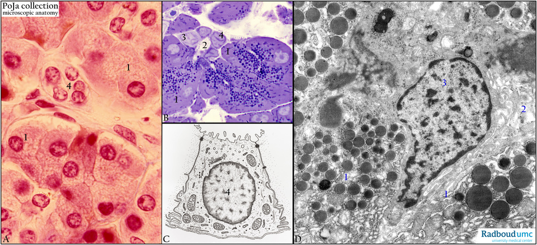
Title: Intercalated duct of exocrine pancreas (human, rabbit)
Description: Stain: (A) Azan. (C) Electron micrograph scheme. (B) Toluidine blue semi-thin section. (D) Electron microscopy.
The acinus consists of secretory serous epithelial cells (1) with apically located granules containing digestive enzymes, which are secreted in the lumen.
In (B, D) lumen (2) of an intercalated duct (4) originating from a centroacinar epithelial cell (3). In contrast to the secretory cells (1), the ductal cells do not contain secretion granules (B), as they produce a bicarbonate solution but no proteins.
Keywords/Mesh: pancreas, exocrine, centroacinar cell, intercalated duct, electron microscopy, histology, POJA collection

