
5.4.3 POJA-L2498+2432+
La0046+2507+2412+2501+
2406+4265+2509+5036.
Tubular system and various protein expressions (XIX) in the kidney
5.4.3 POJA-L2498+2432+La0046+2507+2412+2501+2406+4265+2509+5036
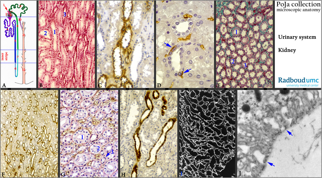
Title: Tubular system and various protein expressions (XIX) in the kidney
Description:
(A): Scheme of nephron, human.
(1) Vascular pole of glomerulus. (7) Ascending thin limb (ATL).
(2) Glomerulus. (8) Distal straight tubule (TAL).
(3) Urinary pole. (9) Distal convoluted tubule (DCT).
(4) Proximal convoluted tubule (PCT). (10) Connective tubule (CNT).
(5) Proximal straight tubule (PST). (11) Cortical collecting duct (CCD).
(6) Descending thin limb (DTL) of the loop of Henle. (12) Medullary collecting duct (IMCD).
(B): Stain Azan, human. (1) Outer medullary collecting duct (OMCD).
(2) Proximal straight tubule (PST).
(3) Medullary thick ascending limb (mTAL) in the outer stripe of outer zone.
(C): Immunoperoxidase staining with DAB and antibodies against Tamm-Horsfall glycoprotein (THP) (uromodelin), human.
The THP is produced by the thick ascending limb (mTAL) of the loop of Henle. OMCD and PST are negative.
(D): Immunoperoxidase staining with DAB and antibodies against Band-3, (AE1) which is highly expressed in intercalated cells
of the convoluted distal tubules (DCT) at the basolateral site (arrows), human.
The red blood cells in the capillaries are also stained (hemoglobin and DAB).
(E): Stain trichrome Goldner, human. Outer stripe of outer zone, with cross-sections through OMCD (1), mTAL (2), PST (3).
(F): Immunoperoxidase staining with DAB and antibodies against THP, expressed in mTAL, inner stripe of outer zone, human.
(G): Stain Weigert hematoxylin-azophloxine, human. Inner stripe of outer zone with OMCD (1), mTAL (2), DTL (3).
(H): Immunoperoxidase staining with DAB and antibodies against calcium-binding protein (CaBP-28 kDa) expressed in cortical
collecting duct (CCD), human. PST is negative.
(I): Immunofluorescence staining with antibodies against heparan sulfate (HSPG), black-white, human. Outer medulla with positive basement membranes and some connective tissue forming an intricate meshwork that fill up the medulla.
(J): Immunogold electron microscopy with antibodies (HS4C3) against heparan sulfate reacting positively (arrows on 10 nm gold
particles) with the basal lamina of a principal cell of the collecting duct (CD), rat.
Keywords/Mesh: urinary system, kidney, collecting duct, OMCD, mTAL, calcium-binding protein, heparan sulfate, Tamm-Horsfall glycoprotein, uromodelin, histology, electron microscopy, POJA collection