THEME-15.0 – THE LOCOMOTOR SYSTEM – CARTILAGE: INTRODUCTION
The locomotor system or Musculo-skeletal system can be considered as an organ system and is made up of:
- POJA-14.0 Locomotor System: Muscles
- POJA-15.0 Locomotor System: Cartilage
- POJA-16.0 Locomotor System: Bone
POJA-15 The Locomotor system – Cartilage chapter is composed of :
- 15.0 Cartilage: Introduction
- 15.1 Hyaline cartilage
- 15.2 Elastic cartilage
- 15.3 Fibrocartilage
- 15.4 Cartilage: Literature References
15.0 Introduction:
Cartilage is an avascular dense connective tissue which consists of cells (chondrocytes) and a solid extracellular matrix (a.o. chondroitin sulphate), that functions as support and protection. Glycosaminoglycans and type II collagen fibres facilitates diffusion of substances between the surrounding vascular connective tissue and the dispersed chondrocytes.
On the basis of the amount and characteristics of the extracellular matrix (ECM) and the prevalent collagenous and elastic fibres embedded in it, light microscopically three kinds of cartilage are distinguished (see below).
Cartilage is avascular and aneural. It receives nutrition by diffusion from perichondral capillaries. Nerve fibres are absent.
Cartilage grows by appositional grow around the chondroblast and/or by interstitial growth of the chondrocytes.
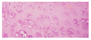
- 15.1 Hyaline cartilage (glassy appearance) is the most common form in the mature body. Its extracellular matrix is made up of type II collagen fibres together with glycosaminoglycans, proteoglycans and multi-adhesive glycoproteins. Hyaline is a precursor for bone and is found in the epiphyseal growth plates of children. It is also found in the ribs, nose, larynx and trachea.
- 15.2 Elastic cartilage (yellow appearance) contains elastic fibres (elastin) in the extracellular matrix (ECM), and also collagen fibres. It is found in the pinnae (the external ear flaps of many mammals, including humans) and in the epiglottis. Chondrocytes lie between the fibres.
- 15.3 Fibrocartilage characterises by the great amount of collagen type I. It is found e.g. in the intervertebral discs and pubic symphysis.
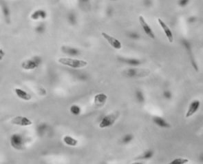
- Foetal cartilage should be considered as a hyaline cartilage although its appearance differs from that in the adult stages as demonstrated below in the black and white image:
|
POJA-L7142 In foetal cartilage blood vessels are found localised. The numerous cartilage cells are spindle-shaped or rounded cells, and even star-like cells are more or less evenly distributed without formation of nests (groups) composed of isogenous cells that is characteristic for mature hyaline cartilage. |


