
14.3 POJA-L6307+6308 Electron micrograph of branching myocytes in the corpus spongiosum of the penis II (human)
14.3 POJA-L6307+6308 Branching myocytes in the corpus spongiosum of the penis II
14.3 POJA-L6307+6308 Electron micrograph of branching myocytes in the corpus spongiosum of the penis II (human)
Title: Electron micrograph of branching myocytes in the corpus spongiosum of the penis II (human)
Description:
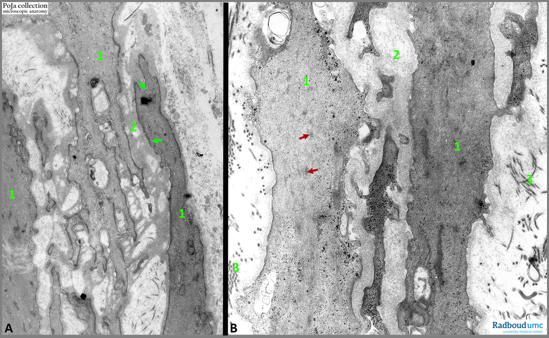
Basically, the corpus spongiosum of the urethra-seminal duct in the penis is composed of cavernous spaces formed by fibro-collagenous stroma supported by branched smooth muscle cells (1) that regulate the blood flow in the cisternal spaces. (2): Close relation of distinct basal lamina with collagen fibrils in the ECM. Note also in (A) the numerous small caveolar structures along the cell membrane (A: green arrows).
Many myocytes possess multiple processes (A, B). Their cytoplasm is studded with actin filaments. Spots of dense plaques (B: red arrows) are found dispersed. (3) Collagen fibres.
Mostly the myocytes are longitudinally organised. The myocytes are also arranged in the so-called subendothelial cushings beneath the endothelia of the vascular channels (or cisterns).
These myocytes regulate the diameter of the cisternal space analogous to the branched myocytes in the media of elastic arteries.
See also:
Keywords/Mesh: locomotor system, penis, corpus spongiosum, smooth muscle, dense plaque, branching smooth muscle, electron microscopy, POJA collection
Description:
Basically, the corpus spongiosum of the urethra-seminal duct in the penis is composed of cavernous spaces formed by fibro-collagenous stroma supported by branched smooth muscle cells (1) that regulate the blood flow in the cisternal spaces. (2): Close relation of distinct basal lamina with collagen fibrils in the ECM. Note also in (A) the numerous small caveolar structures along the cell membrane (A: green arrows).
Many myocytes possess multiple processes (A, B). Their cytoplasm is studded with actin filaments. Spots of dense plaques (B: red arrows) are found dispersed. (3) Collagen fibres.
Mostly the myocytes are longitudinally organised. The myocytes are also arranged in the so-called subendothelial cushings beneath the endothelia of the vascular channels (or cisterns).
These myocytes regulate the diameter of the cisternal space analogous to the branched myocytes in the media of elastic arteries.
See also:
- 13.1 POJA-L4719+4594+4561+4564+4596+4563+4565+4572 Elastic artery: light and electron microscopy
- 14.3 POJA-L6314+6315 Smooth muscles in corpus spongiosum of the penis I
Keywords/Mesh: locomotor system, penis, corpus spongiosum, smooth muscle, dense plaque, branching smooth muscle, electron microscopy, POJA collection