
4.2.1 POJA-L3754+3767. Keratin 7 staining of intrahepatic bile duct and terminal bile ductule (Hering) (human)
4.2.1 POJA-L3754+3767
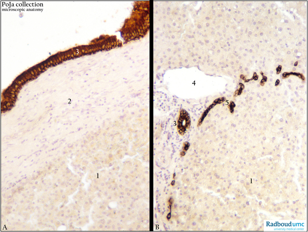
Title: Keratin 7 staining of intrahepatic bile duct and terminal bile ductule (Hering) (human)
Description: Immunoperoxidase-DAB staining with anti-cytokeratin 7 (OVTL12/30 antibodies).
(A) Intrahepatic bile duct.
(1) Unstained liver parenchym cells. (A, 2) Connective tissue liver capsule. (A, 3) Intrahepatic bile duct epithelium stained positively with cytokeratin 7.
(B) Ductuli of Hering.
(B, 3) Portal bile canaliculus or ductules (CK-7 positive). (B, 5) Hering ductules are CK7 positive. Note that as soon as the liver parenchymal cells turn into canaliculi cell types they begin to express cytokeratin 7. (B, 5) Branch of the portal vein.
Keywords/Mesh: liver cells, ductule of Hering, bile duct, histology, POJA collection