
13.1 POJA-L2979+4643+4644+4692+4334 Ultrastructure of capillaries
13.1 POJA-L2979+4643+4644+4692+4334
Title: Ultrastructure of capillaries
Description:
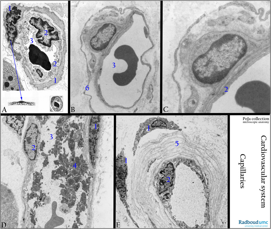
(A): Electron micrograph scheme of a capillary. The capillary wall is composed of endothelial cells (2), a basal lamina and few contractile cells i.e. the pericytes (1). (3) Lumen capillary. The endothelial cells are anchored to each other.
(B, C): Electron micrographs of capillaries from the region of the main bronchus (golden hamster) with similar structures. Note that there is a common basal lamina between the outer located pericyte [(1) in the scheme] and the endothelium (2). Long slender processes of a perivascular fibroblast (also called interstitial fibroblast) (6) and thin nerve fibres.
(D): The venous capillary in is filled with aggregates of platelets (4), which are closely adhered to the endothelial lining. (electron micrograph, exocrine pancreas, dog). (2) Endothelial nucleus, (1) pericyte nucleus. Perivascular oedema is present outside the capillary.
(E): Electron micrograph of a capillary with multiple basal laminae (mucosa of lip region), human, PXE (see under Background). Distinct multiplication of basal lamina layers (5) and (1) perivascular fibroblasts, circle around capillary lumen. Tip of pericytic process between the basal laminae. In several disorders (i.a. diabetes, cutaneous lesions in lupus erythematosus) capillaries are also frequently observed to be enveloped with layers of basal laminae.
Background: Gap junctions are present between the endothelial cells and pericytes of the capillaries. These pericytes line the outer surface of blood vessels and stabilise the vessel walls. They are contractile cells that control the blood circulation in capillaries and post-capillary venules. After tissue damage pericytes can transform into smooth muscle cells of arterioles and venules during neo-angiogenesis.
Pseudoxanthoma elasticum (PXE) is a disorder characterised by progressive calcification and fragmentation of elastic fibres, and cutaneous lesions. PXE involves elastic fibres i.a. in the reticular dermis of the skin and in smaller/mid-sized arteries. Ogawa, K. (1981), Ultrastructure of cutaneous lesions in lupus erythematosus: a comparison between the cutaneous and systemic types. The Journal of Dermatology, 8: 175–186. doi: 10.1111/j.1346-8138.1981.tb02532.x
Keywords/Mesh: cardiovascular system, vascularisation, blood vessel, capillary, endothelium, basal lamina, pericyte, fibroblast, Pseudoxanthoma elasticum, electron microscopy, histology, POJA collection
Title: Ultrastructure of capillaries
Description:
(A): Electron micrograph scheme of a capillary. The capillary wall is composed of endothelial cells (2), a basal lamina and few contractile cells i.e. the pericytes (1). (3) Lumen capillary. The endothelial cells are anchored to each other.
(B, C): Electron micrographs of capillaries from the region of the main bronchus (golden hamster) with similar structures. Note that there is a common basal lamina between the outer located pericyte [(1) in the scheme] and the endothelium (2). Long slender processes of a perivascular fibroblast (also called interstitial fibroblast) (6) and thin nerve fibres.
(D): The venous capillary in is filled with aggregates of platelets (4), which are closely adhered to the endothelial lining. (electron micrograph, exocrine pancreas, dog). (2) Endothelial nucleus, (1) pericyte nucleus. Perivascular oedema is present outside the capillary.
(E): Electron micrograph of a capillary with multiple basal laminae (mucosa of lip region), human, PXE (see under Background). Distinct multiplication of basal lamina layers (5) and (1) perivascular fibroblasts, circle around capillary lumen. Tip of pericytic process between the basal laminae. In several disorders (i.a. diabetes, cutaneous lesions in lupus erythematosus) capillaries are also frequently observed to be enveloped with layers of basal laminae.
Background: Gap junctions are present between the endothelial cells and pericytes of the capillaries. These pericytes line the outer surface of blood vessels and stabilise the vessel walls. They are contractile cells that control the blood circulation in capillaries and post-capillary venules. After tissue damage pericytes can transform into smooth muscle cells of arterioles and venules during neo-angiogenesis.
Pseudoxanthoma elasticum (PXE) is a disorder characterised by progressive calcification and fragmentation of elastic fibres, and cutaneous lesions. PXE involves elastic fibres i.a. in the reticular dermis of the skin and in smaller/mid-sized arteries. Ogawa, K. (1981), Ultrastructure of cutaneous lesions in lupus erythematosus: a comparison between the cutaneous and systemic types. The Journal of Dermatology, 8: 175–186. doi: 10.1111/j.1346-8138.1981.tb02532.x
Keywords/Mesh: cardiovascular system, vascularisation, blood vessel, capillary, endothelium, basal lamina, pericyte, fibroblast, Pseudoxanthoma elasticum, electron microscopy, histology, POJA collection