12.1.4 POJA-L2571+2577+2567+4424
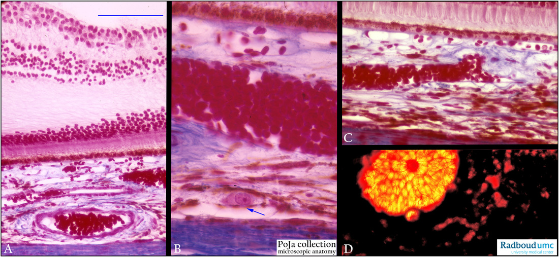
Title: Choroid and fovea centralis in retina
Description:
(A, B, C): Retina with fovea centralis, stain Azan, human. (A) Indicates the parafoveal area near to the inner centre the foveola. The choroid is supplied by a network of vessels from the long ciliary artery (a branch from the ophthalmic artery). Branches of these vessels form an intricate network of capillaries i.e. the choriocapillaris close to pigmented epithelium (RPE). The choroid below the area of the fovea centralis shows the choriocapillaris of the choroid layer adjacent to the pigmented epithelial cell layer (RPE) of the retina and also locally well-developed blood vessels (in A) that supply oxygen specifically to this area.
The choroidal stroma is a connective tissue layer with blood vessels, nerves, few smooth muscles and melanocytes. The stroma adheres to the inner pigmented layer of the sclera, the so-called lamina fusca.
Note in (B) a solitary ganglion cell (arrow) as well as brown melanised melanocytes in the choroid and the pigmented lamina fusca.
(D): Uvea, in situ perfusion with Sirius red, human.The uvea or the pigmented vascularised middle tunic is divided into the choroid, ciliary body and iris. In situ perfusion of the uvea shows reddish-stained spots of choroidal venous branches close to the bright yellow-red orifice of the formerly optic disc.
Keywords/Mesh: eye, retina, fovea centralis, choroid, uvea, histology, POJA collection

