7.2 POJA-L1552+1553
Title: Macroscopy leiomyomas (uterus, human).
Description:
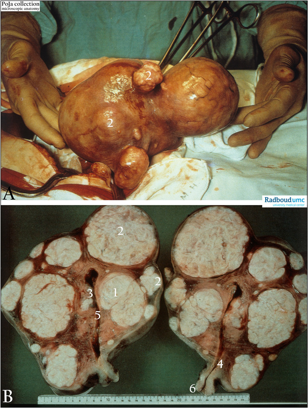
(A): Uterine myomatosis after abdominal hysterectomy.
(B): Median section of leiomyomas in uterus with (1) intramural, (2) subserosal and (3) submucosal localisations ; (4) cervix; (5) uterine cavity; (6) vagina.
The myomas appear round sometimes reaching up to 20 cm or more in diameter. The white solid but elastic mass consists of whorly bundles of smooth muscle cells and fibers separated by connective tissue stroma. The nodules can be hyalinised and calcified and these degenerations are composed of homogenous plaques, microscopically equivalent to eosinophilic zones without any cells between the smooth muscle cell bundles. (By courtesy of G. P. Vooijs MD PhD, former Head of the Department of Pathology, and the Museum of Anatomy and Pathology, Radboud university medical center, Nijmegen, The Netherlands).
Clinical background: Leiomyoma (myoma uteri): A leiomyoma is benign tumor derived from the smooth muscle cells from the myometrium accompagnied by connective tissue structures (therefore also called uterine myomatosis or fibromyoma). These tumors are frequently encountered in 35 year over older women, and often occur in multiple forms. The tumor increases in size during gravidity as well as during estrogen induced anti-conception period. Patients with myomas usually do have higher serum levels of growth hormone and estradiol. Myoma can be removed by enucleatie or by complete hysterectomy. Myomas are classified macroscopically according to localization: the intramural myoma is most common and is localised in the uterine corps; the subserosal myoma is mostly pedunculated and is localized subperitoneally; the submucosal myoma is covered by the endometrium and bulges into the uterine cavity. If this tumor is pedunculated it can be classified as well as myomatous polyps; the intraligamentous myoma grows out from the uterine wall in between the blades of the ligamentum latum (broad ligament).
Keywords/Mesh: female reproductive organs, uterus, leiomyoma, macroscopy, myomatosis, cervix, vagina, histology, POJA collection.

