
5.4.3 POJA-L2396+2436+
5002+2393+2478+2480+
La0352.
Proximal tubules (XI) in kidney
5.4.3 POJA-L2396+2436+5002+2393+2478+2480+La0352
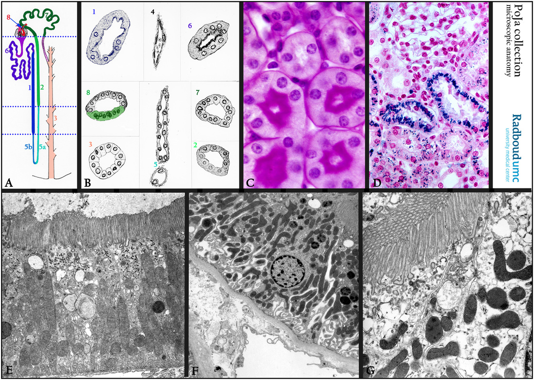
Title: Proximal tubules (XI) in kidney
Description:
(A, B): Scheme nephron, human. (1) Pars recta I of the proximal tubule or thick descending limb or proximal straight tubule (PST).
(2) Pars recta II or thick ascending limb (TAL).
(3) Collecting duct or tubule.
(4) Capillary.
(5) Henle’s loop comprising the thin ascending limb (5a) and the thin descending limb (5b).
(6) Pars contorta I or proximal convoluted tubule (PCT).
(7) Pars contorta II or distal convoluted tubule (DCT).
(8) Macula densa as part of the cortical thick limb of Henle (cTALH).
(C): Kidney cortex, stain PAS, mouse. Both the microvilli of the proximal tubules and the basal lamina of the cells are stained positively.
(D): Kidney cortex, stain modified hematoylin-eosin, mouse.
Uptake of injected trypan blue by the cells of the PCT (proximal convoluted tubules). The PST (proximal straight tubule) absorbs
much less trypan blue.
(E): Electron microscopy, rabbit, proximal straight tubule. Note long and regular microvilli and the slender apical endocytotic vesicles, basally long mitochondria and several lysosomal structures.
(E, by courtesy of A. Stadhouders PhD, former head Department Electron Microscopy, University Medical Centre of the Radboud University, Nijmegen, the Netherlands).
(F): Electron microscopy, PCT(proximal convoluted tubule), human. Note the basal invaginations and the enfolded mitochondria.
(G): Electron micrograph of the apex of a PCT cell, human. Note the extended microvilli between which the endocytotic vesicles are pinched off. Both the microvilli (brush border) and the numerous mitochondria are needed for active transport of sodium ions out of
the cells to create a concentration gradient which allows more sodium ions to enter the cell from the luminal side.
Water passively follows the sodium out of the cell along its concentration gradient. (Na+/K+ ATPase in the basolateral membrane).
The distal tubules do not have an apical brush border, are less eosinophilic and they are involved in the ATP-dependent regulation of calcium (and Na/K).
Keywords/Mesh: urinary system, kidney, proximal tubules, distal tubules, histology, electron microscopy, POJA collection