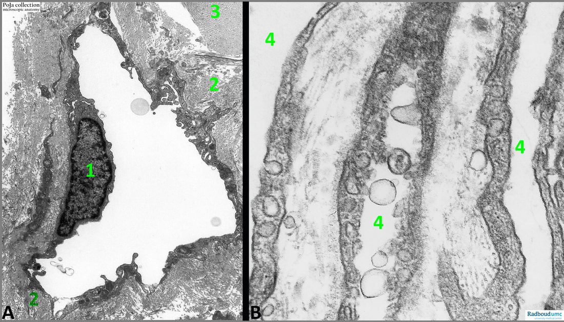
14.1 POJA-L6312+6313 Electron micrograph of lymph capillaries in skeletal muscle (human)
14.1 POJA-L6312+6313 Electron micrograph of lymph capillaries in skeletal muscle (human)
Title: Electron micrograph of lymph capillaries in skeletal muscle (human)
Description:
(A): Electron micrograph of a lymph capillary in muscular tissue: it has extremely thin walls consisting of one layer of lymphatic endothelial cells (1) without a basal membrane. Instead, there is a layer of fibrils (2) (collagen IV and VI) underneath the endothelium included some cross anchoring filaments to connect connective tissue with the endothelium. (3): Muscle fibre filaments.
(B): In the perimysium these longitudinal sectioned capillar tubes or fissures (4: lumen) collect the tissue liquid. They are partly fenestrated and function as drainage system. The lining endothelial cells present many pinocytotic vesicles (or caveolae) but no basal lamina.
See also:
- 13.1 POJA-La0320+L-4672 Lymphatic capillaries
Keywords/Mesh: locomotor system, striated muscle, skeletal muscle, vascularisation, lymph vessel, lymph capillary, pinocytosis, basal lamina, electron microscopy, POJA collection

