
7.4 POJA-L-105+102. Mammary gland secretion (human, monkey)
7.4 POJA-L-105+102
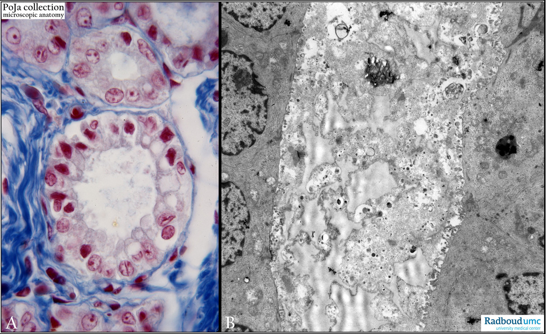
Title: Mammary gland secretion (human, monkey)
Description: Stain: (A) Azan (human); (B) Electron microscopy (monkey).
(A): Acini (alveoli) filled with secretion and intracellular secretion vacuoles in glandular epithelium.
(B): Lumen of an acinus (alveolus) filled with secretion that consists of a mixture of heterogeneous structures such as remnants of cytoplasmic structures. Smaller electron-dense spots could represent proteinaceous products. Adjoining epithelial cells show short apical microvilli.
Background: The mammary gland is classified as compound tubule-alveolar gland. Milk contains proteins that will be discharged by merocrine secretion (released secretory vesicles) while lipids (triglycerides, cholesterol) are released by the apocrine mode of secretion. Prolactin (from the pituitary gland) stimulates secretion by alveolar cells. Oxytocin stimulates the contraction of myoepithelial cells causing the ejection of milk.
Keyword(s)/Mesh: breast, mammary glands, lactation, histology, electron microscopy, POJA collection