
11.5 POJA-L3371+3147+
2998+3001+3359.
Fetal and adult cerebrum
11.5 POJA-L3371+3147+2998+3001+3359
Title: Fetal and adult cerebrum
Description:
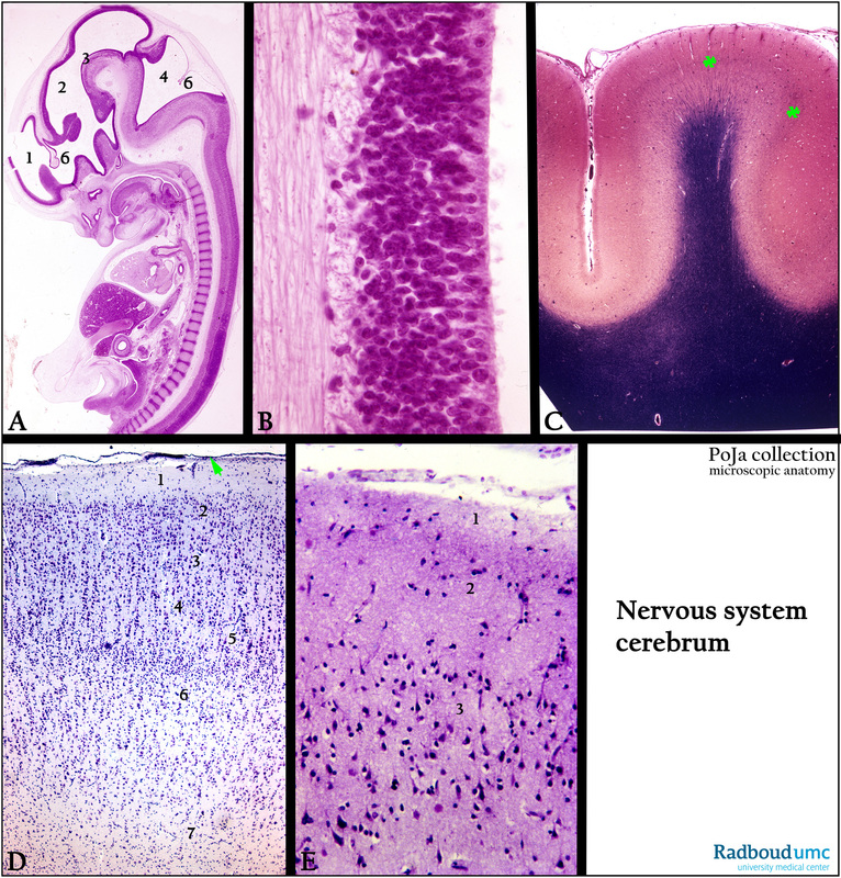
(A): Stain hematoxylin-eosin, fetus (ca. 3 months), human.
(1) Telencephalon/forebrain. (4) 4th ventricle with roof of rhombencephalon/hindbrain.
(2) Mesencephalon/midbrain. (6) Choroid plexus.
(3) Cephalic flexure (midbrain).
(B): Stain hematoxylin-eosin, fetus (less than 3 months), human. The ventricular zone of the wall of the neural tube in the early development.
(C): Stain modified Weigert, human. The myelin in the medulla is stained black, (green **). External band of Baillager is visible in the cortex and note the pia mater at the surface.
(D): Stain cresyl violet, human, illustrating the general structure plan of the cortex of the cerebrum (associative area). Pia mater (green arrow).
(1) Lamina zonalis or molecularis (I. Molecular layer).
(2) Lamina granularis externa (II. External granular layer).
(3) Lamina pyramidalis externa (III. External pyramidal layer).
(4) Lamina granularis interna (IV. Internal granular layer).
(5) Lamina ganglionaris (V. Internal pyramidal layer).
(6) Lamina multiformis (VI. Multiform layer).
(E): Stain cresyl violet, human. Shapes of the different neurons in the external layers in the associative cortex.
(1) Molecular layer.
(2) External granular layer.
(3) External pyramidal layer.
Keywords/Mesh: nervous tissue, fetus, cerebrum, isocortex, laminar architecture, histology, POJA collection
Title: Fetal and adult cerebrum
Description:
(A): Stain hematoxylin-eosin, fetus (ca. 3 months), human.
(1) Telencephalon/forebrain. (4) 4th ventricle with roof of rhombencephalon/hindbrain.
(2) Mesencephalon/midbrain. (6) Choroid plexus.
(3) Cephalic flexure (midbrain).
(B): Stain hematoxylin-eosin, fetus (less than 3 months), human. The ventricular zone of the wall of the neural tube in the early development.
(C): Stain modified Weigert, human. The myelin in the medulla is stained black, (green **). External band of Baillager is visible in the cortex and note the pia mater at the surface.
(D): Stain cresyl violet, human, illustrating the general structure plan of the cortex of the cerebrum (associative area). Pia mater (green arrow).
(1) Lamina zonalis or molecularis (I. Molecular layer).
(2) Lamina granularis externa (II. External granular layer).
(3) Lamina pyramidalis externa (III. External pyramidal layer).
(4) Lamina granularis interna (IV. Internal granular layer).
(5) Lamina ganglionaris (V. Internal pyramidal layer).
(6) Lamina multiformis (VI. Multiform layer).
(E): Stain cresyl violet, human. Shapes of the different neurons in the external layers in the associative cortex.
(1) Molecular layer.
(2) External granular layer.
(3) External pyramidal layer.
Keywords/Mesh: nervous tissue, fetus, cerebrum, isocortex, laminar architecture, histology, POJA collection