
11.2.2 POJA-L3294+3284+
3286+3287+3285+3288+
3289.
Sensory receptors (neuromuscular spindles)
11.2.2 POJA-L3294+3284+3286+3287+3285+3288+3289
Title: Sensory receptors (neuromuscular spindles)
Description:
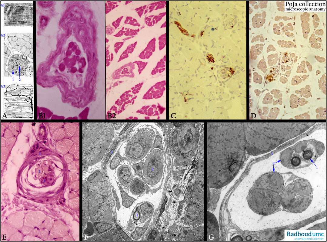
(A): Scheme striated muscle with neuromuscular spindle (afferent sensory receptor), human. (A1) Longitudinal section of striated muscle fibers with A- bands, I-bands and Z-lines, endomysium and fiber cell nuclei. (A2) Cross-sectioned striated muscle with neuromuscular spindle. (1) Myelinated axonal terminals within the muscle spindle. (2) Four to twelve intrafusal muscle fibers with a centrally located nucleus, which are innervated separately from the striated muscles. (A3) Capillary network in the striated muscle running parallel to the longitudinal sectioned muscle fibers and engulf each individual fiber as well (Indian ink perfusion).
Background 1: The intrafusal muscle fibers register changes in tonus and length within the muscle. The spindle is surrounded by a connective tissue capsule and the entire interior is filled with liquid. With contraction and relaxation of striated (extrafusal) muscle fibers the proprioreceptors are stimulated. Afferent nerves ends on the intrafusal fibers of
the neuromuscular spindle and react on contraction of the large extrafusal fibers outside of the spindle. Additonally an efferent innervations of the intrafusal fibers is also present.
(B 1, 2): Stain hematoxylin-eosin, lumbricals of hand, human. Survey and detail of the neuromuscular spindles within the striated muscle bundle, equivalent to (A2).
(C): Immunoperoxidase staining with AEC and antibodies against neurofilament, 4d postnatal rat. The presence of the neuromuscular spindles in the striated muscles in the dermis is illustrated. Note only the axons running toward muscle spindles as well as the presence of the spindle’s afferent/efferent endings.
(D): Immunoperoxidase staining with DAB and antibodies against the intermediate filament GFAP (glial fibrillary acidic protein), 4d postnatal rat. It illustrates the presence of the spindles in the striated muscles of the dermis. Myelinated nerves and Schwann cells are found within or close by the spindles.
(E): Stain hematoxylin-eosin, lumbricals of hand, human. Detail of a muscle spindle corresponding to the electron micrographs of those in the soleus muscle in (F, G).
(F): Electron micrograph of muscle spindle, mouse. (1) Sensory axons. (2) Intrafusal muscle fibers. (3) Connective tissue capsule as an extension of the perineurium.
(G): Electron micrograph of muscle spindle, mouse. (1) Myelinated sensory axons, (2) intrafusal muscle fibers, and (3) connective tissue fibers as part of the surrounding capsule, mouse.
Background 2: Two types of intrafusal muscle fibers are known: the nuclear bag fibers and the nuclear chain fibers. The nuclei are concentrated in the expanded bag-like equatorial region, i.e. the nuclear bag. The nuclear chain fibers are thicker and longer and are lined-up in the equatorial region-the nuclear chain.
Keywords/Mesh: nervous tissue, proprioreceptor, muscle receptor, muscle spindle, neuromuscular spindle, striated muscle, histology, electron microscopy, POJA collection
Title: Sensory receptors (neuromuscular spindles)
Description:
(A): Scheme striated muscle with neuromuscular spindle (afferent sensory receptor), human. (A1) Longitudinal section of striated muscle fibers with A- bands, I-bands and Z-lines, endomysium and fiber cell nuclei. (A2) Cross-sectioned striated muscle with neuromuscular spindle. (1) Myelinated axonal terminals within the muscle spindle. (2) Four to twelve intrafusal muscle fibers with a centrally located nucleus, which are innervated separately from the striated muscles. (A3) Capillary network in the striated muscle running parallel to the longitudinal sectioned muscle fibers and engulf each individual fiber as well (Indian ink perfusion).
Background 1: The intrafusal muscle fibers register changes in tonus and length within the muscle. The spindle is surrounded by a connective tissue capsule and the entire interior is filled with liquid. With contraction and relaxation of striated (extrafusal) muscle fibers the proprioreceptors are stimulated. Afferent nerves ends on the intrafusal fibers of
the neuromuscular spindle and react on contraction of the large extrafusal fibers outside of the spindle. Additonally an efferent innervations of the intrafusal fibers is also present.
(B 1, 2): Stain hematoxylin-eosin, lumbricals of hand, human. Survey and detail of the neuromuscular spindles within the striated muscle bundle, equivalent to (A2).
(C): Immunoperoxidase staining with AEC and antibodies against neurofilament, 4d postnatal rat. The presence of the neuromuscular spindles in the striated muscles in the dermis is illustrated. Note only the axons running toward muscle spindles as well as the presence of the spindle’s afferent/efferent endings.
(D): Immunoperoxidase staining with DAB and antibodies against the intermediate filament GFAP (glial fibrillary acidic protein), 4d postnatal rat. It illustrates the presence of the spindles in the striated muscles of the dermis. Myelinated nerves and Schwann cells are found within or close by the spindles.
(E): Stain hematoxylin-eosin, lumbricals of hand, human. Detail of a muscle spindle corresponding to the electron micrographs of those in the soleus muscle in (F, G).
(F): Electron micrograph of muscle spindle, mouse. (1) Sensory axons. (2) Intrafusal muscle fibers. (3) Connective tissue capsule as an extension of the perineurium.
(G): Electron micrograph of muscle spindle, mouse. (1) Myelinated sensory axons, (2) intrafusal muscle fibers, and (3) connective tissue fibers as part of the surrounding capsule, mouse.
Background 2: Two types of intrafusal muscle fibers are known: the nuclear bag fibers and the nuclear chain fibers. The nuclei are concentrated in the expanded bag-like equatorial region, i.e. the nuclear bag. The nuclear chain fibers are thicker and longer and are lined-up in the equatorial region-the nuclear chain.
Keywords/Mesh: nervous tissue, proprioreceptor, muscle receptor, muscle spindle, neuromuscular spindle, striated muscle, histology, electron microscopy, POJA collection