
13.1 POJA-L4700+4701+4702+4703+4704 Dermal nodules in Kaposi sarcoma
13.1 POJA-L4700+4701+4702+4703+4704
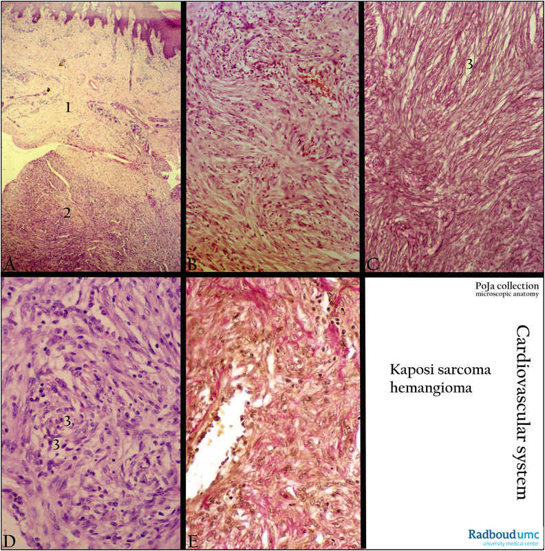
Title: Kaposi sarcoma (human)
Description:
(A, B, D): Haematoxylin-eosin staining. (C). Silver stain (Laguesse). (E). van Gieson. Poorly defined dermal nodules (2) or plaques in papillary dermis and subcutis (1) consisting of spindle cells. The van Gieson stain in (E) shows redly stained elastic fibres and brown stained collagen fibres.
At medium magnification clefts and ovoid spaces (3) lined with elongated endothelial cells that are either empty or filled with erythrocytes. In addition, there are solid sprouts of endothelial-like cells without canalisation, intermingled with reticular fibres (in C).
Background: Kaposi sarcoma is a low-grade vascular tumor (malignant haemangioendothelioma) that may involve the skin, oral mucosa, lymph nodes and visceral organs. It is composed of vascular endothelium and associated with Kaposi sarcoma herpesvirus. The classic Kaposi sarcoma has a more indolent course than the AIDS-related sarcoma. It progresses over 10-15 years with gradual enlargement of cutaneous lesions. At present it is a prevalent malignancy among patients with HIV, drug-related or transplant-associated immunosuppression and has an aggressive clinical course.
(Oana Radu and Liron Pantanowitz (2013) Kaposi Sarcoma. Archives of Pathology & Laboratory Medicine: February 2013, vol 137, No 2, pp 289-294). Doi: http://dx.doi.org/10.5858/arpa.2012-0101-RS
Keywords/Mesh: cardiovascular system, vascularisation, blood vessel, capillary, vascular tumor, haemangioendothelioma, Kaposi sarcoma, AIDS, histology, POJA collection