7.1 POJA-L1502+1842
Title: Antral follicles, ovary (human)
Description: Stain: (A, B) Hematoxylin-eosin.
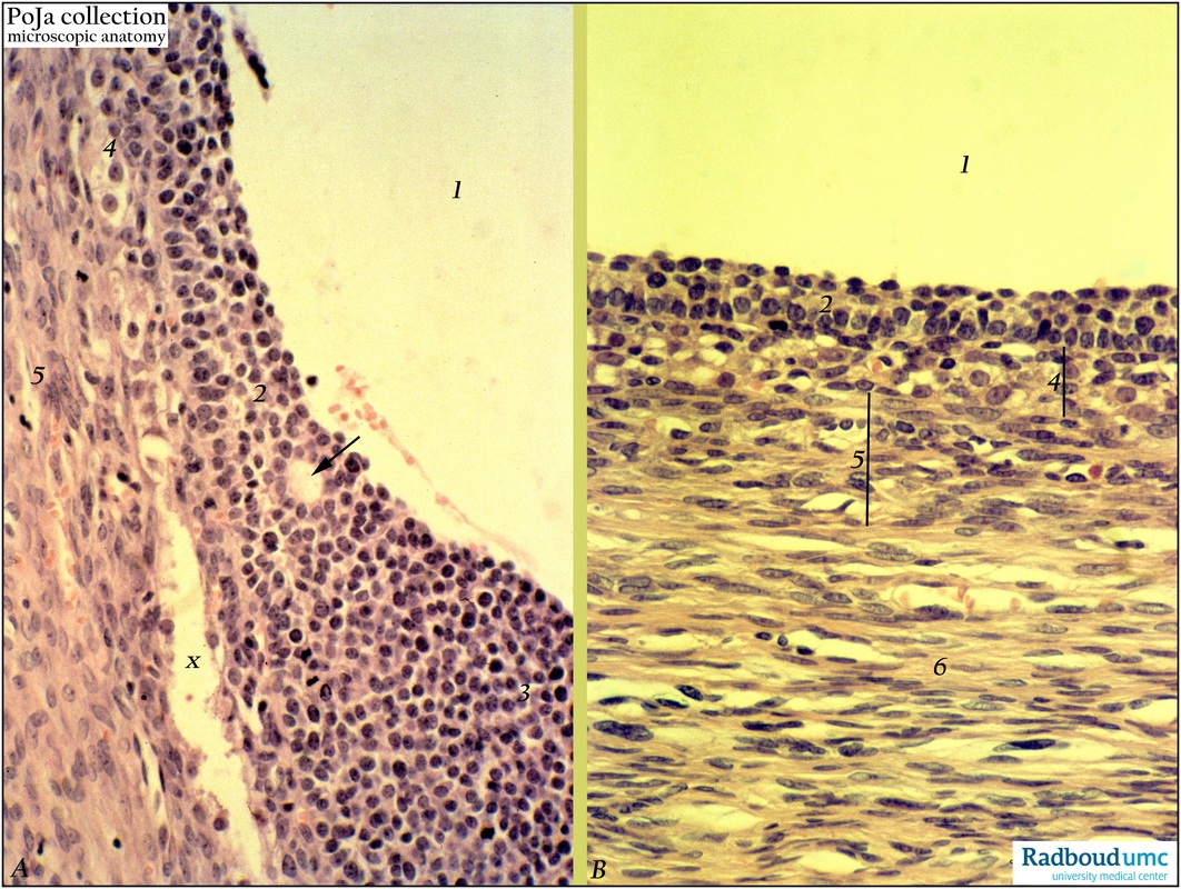
(A): Large normal antral follicle (1) type 5+. Transition of well-formed mural granulosa layer (2, membrana granulosa) into cumulus oophorus (cumulus ovaricus) (3). At (arrow) an occasional Call-Exner body (small intercellular space filled with follicular fluid). Close to the granulosa cells vascularized internal theca cells (4) followed by a more fibrous layer of externa theca cells (5). (At x technical preparation artifact).
(B): Large antrum (1) of a follicular cyst, the wall is lined by layers of almost compressed mural granulosa cells (2) and an inner/outer layer of theca (4, 5) with small blood vessels followed by increased fibrous layers (6). The follicle grows larger than normal and does not rupture to release the oocyte.
Background: In normal antral follicles the follicular fluid is semi-viscous, yellow-colored complex extracellular fluid. It is derived mainly from plasma via the vascular compartment in the follicle wall. The largest osmotically active molecules are chondroitin sulphate proteoglycans, hyaluronan as well as versican and inter-alpha trypsin inhibitor. Follicular cysts are usually single, thin-walled and unilocular; they can occur at any age and might range from 3-10 cm. Most follicular cysts are asymptomatic but may occasionally rupture and cause a hemoperitoneum. Some appear to be estrogenic and are associated with menstrual disturbances.
Keywords/Mesh: female reproductive organs, ovary, tertiary follicles, female genitalia, follicular cysts, antral follicles, Call-Exner bodies, cumulus oophorus, histology, POJA collection

