7.1 POJA-L1330+1647+1904
Title: Corpus luteum in ovary (human)
Description: Stain: (A, B) Hematoxylin-eosin; (C) Scheme electron microscopy.
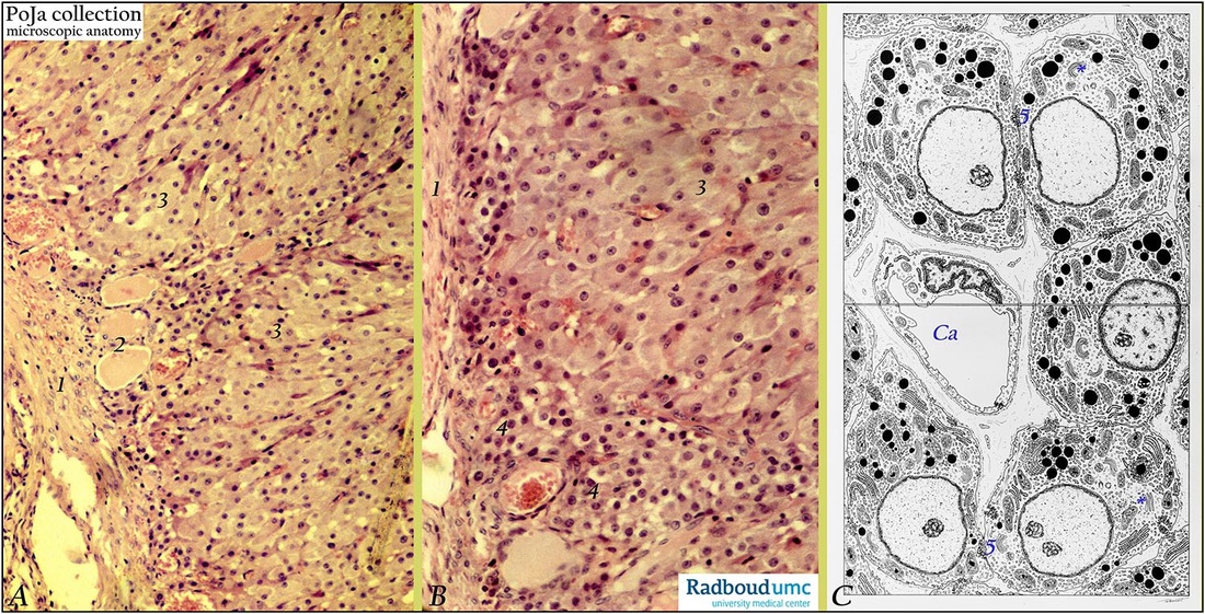
(A): Fibrous border (1) of corpus luteum of pregnancy, infolding with septum and blood vessels (2). Epithelioid arranged sheets of polygonal granulosa lutein cells (3).
(B): Larger granulosa lutein cells (3). Group of smaller theca lutein cells (or paralutein cells) with smaller round nuclei (4) localized peripheral with blood vessels. They also store lipid droplets. Note that the granulosa lutein cells do have larger nuclei.
(C): Scheme: Top side luteinization of granulosa cells, attached by local interdigitations (5). Electron-dense lipid droplets, tubular mitochondria, few Golgi stacks (*) and rough endoplasmic reticulum are present. Vesicular smooth endoplasmic reticulum is abundant. Bottom side drawing shows granulosa lutein cells with increased synthetic activity (more euchromatin in nucleus). There are less interdigitated attachments (5), increased amounts of Golgi areas (*) closely associated with production of new lipid droplets (darkly stained). There are more rough endoplasmic reticulum and enlarged mitochondria. Capillary at (Ca).
Background: After ovulation the lining of granulosa cells of the empty follicle collapses, becomes folded. Under influence of luteinizing hormone (LH) the luteinization process starts i.e. the granulosa cells increase in size and differentiate into granulosa lutein cells while they produce a yellow carotenoid pigment (lutein) as well as progesterone/estrogen. The basal membrane of the collapsed follicle is broken down and internal theca cells invade into the cellular mass together with capillaries and connective tissue. The theca interna cells differentiate into theca lutein cells (or paralutein cells). Together with the granulosa lutein cells, they form epithelioid sheets of round or polygonal lutein cells with fat droplets together constituting the parenchyma of a corpus luteum. The accompanying external theca cells provide stroma and septa of the enlarging corpus luteum that is supplied by numerous blood vessels.
Keywords/Mesh: female reproductive organs, luteal cells, granulosa cells, theca cells,female genitalia, ovary, corpus luteum, histology, POJA collection

