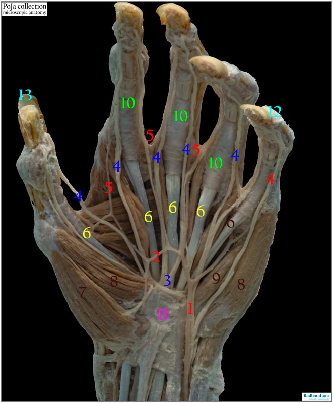
14.5 POJA-L6355 Anatomy of the hand: tendons and muscles
14.5 POJA-L6355 Anatomy of the hand: tendons and muscles
(By courtesy of J. Kooloos PhD and L. Boer PhD, Department Medical Imaging, Anatomy, Museum for Anatomy and Pathology,, Radboud university medical center, Nijmegen The Netherlands)
Title: Anatomy of the hand: tendons and muscles
Description:
Left hand: Arteries are flat, thin and lightly brown while nerves are more compact, massive white-yellow threads.
(1): Superficial branch of ulnar artery
(2): Superficial palmar arch as common stem of ulnar artery with artery to the thumb (left side) and palmar digital arteries to the digits (top side)
(3): Median nerve (dividing into palmar digital branches)
(4): Palmar digital nerve
(5): Palmar digital artery
(6): Flexor digitorum superficialis (yellow)
(7): Abductor pollicis brevis
(8): (left site): Flexor pollicis brevis
(8): (right site): Abductor digiti minimi
(9): Flexor digiti minimi brevis
(10): Fibrous flexor sheath or annulare and cruciforme ligaments
(11): Flexor retinaculum
(I3): Digit I
(12): Digit V
The flexor tendons run from the forearm to the ends of the fingers across the palm side of the hand. These flexors control the bending of fingers down to the palm. A tendon sheath is a thin connective tissue layer that surrounds any tendon. The outer membrane is the outer fibrous tendon sheath (see 10), the inner membrane is the synovial sheath that covers a tendon (e.g. the common synovial sheath for the hand flexors tendons or the ulnar bursa as a synovial sheath in the carpal tunnel of the human hand). Bursae are located where bones, ligaments, tendons and muscles are found and may run together. These bursae are flat fibrous sacs with lining synovial membranes that produce a thin film of synovial fluid. In this way a tendon sheath can be assumed as a lengthened bursae wrapping totally around a tendon subjected to friction.
Dense regular connective tissue is found as the main functional component in:
Tendons attach muscle to bone. The whole tendon (see 6) transmits forces from muscle to bone, it is a non-flexible, strong fibrous structure and serves a.o. to move the bone
Ligaments attach bone to bone and as a flexible, elastic fibrous structure it usually holds structures together and keep them stable. Generally ligaments are similar to tendons but histologically their fibres and the organisation in fascicles is less well ordered. In both tendons as well as ligaments fascicles are separated by an irregular dense connective tissue the endotendineum containing blood vessels and nerves.
Fasciae are similar to ligaments and tendons but surround muscles or other structures. Fascia can be classified as superficial, deep, visceral, or parietal and further classified according to anatomical location.
See also for details:
Keywords/Mesh: locomotor system, muscle, tendon, fascia, ligament, hand, macroscopy, histology, POJA collection.
(By courtesy of J. Kooloos PhD and L. Boer PhD, Department Medical Imaging, Anatomy, Museum for Anatomy and Pathology,, Radboud university medical center, Nijmegen The Netherlands)
Title: Anatomy of the hand: tendons and muscles
Description:
Left hand: Arteries are flat, thin and lightly brown while nerves are more compact, massive white-yellow threads.
(1): Superficial branch of ulnar artery
(2): Superficial palmar arch as common stem of ulnar artery with artery to the thumb (left side) and palmar digital arteries to the digits (top side)
(3): Median nerve (dividing into palmar digital branches)
(4): Palmar digital nerve
(5): Palmar digital artery
(6): Flexor digitorum superficialis (yellow)
(7): Abductor pollicis brevis
(8): (left site): Flexor pollicis brevis
(8): (right site): Abductor digiti minimi
(9): Flexor digiti minimi brevis
(10): Fibrous flexor sheath or annulare and cruciforme ligaments
(11): Flexor retinaculum
(I3): Digit I
(12): Digit V
The flexor tendons run from the forearm to the ends of the fingers across the palm side of the hand. These flexors control the bending of fingers down to the palm. A tendon sheath is a thin connective tissue layer that surrounds any tendon. The outer membrane is the outer fibrous tendon sheath (see 10), the inner membrane is the synovial sheath that covers a tendon (e.g. the common synovial sheath for the hand flexors tendons or the ulnar bursa as a synovial sheath in the carpal tunnel of the human hand). Bursae are located where bones, ligaments, tendons and muscles are found and may run together. These bursae are flat fibrous sacs with lining synovial membranes that produce a thin film of synovial fluid. In this way a tendon sheath can be assumed as a lengthened bursae wrapping totally around a tendon subjected to friction.
Dense regular connective tissue is found as the main functional component in:
- Tendons
- Ligaments
- Fasciae and
- Aponeuroses.
Tendons attach muscle to bone. The whole tendon (see 6) transmits forces from muscle to bone, it is a non-flexible, strong fibrous structure and serves a.o. to move the bone
Ligaments attach bone to bone and as a flexible, elastic fibrous structure it usually holds structures together and keep them stable. Generally ligaments are similar to tendons but histologically their fibres and the organisation in fascicles is less well ordered. In both tendons as well as ligaments fascicles are separated by an irregular dense connective tissue the endotendineum containing blood vessels and nerves.
Fasciae are similar to ligaments and tendons but surround muscles or other structures. Fascia can be classified as superficial, deep, visceral, or parietal and further classified according to anatomical location.
- A retinaculum (see 11) is found in the knee, foot and hand and covers tendons and nerves crossing the joints. It is a fibrous band of a thick fascia around tendons that holds them in place as well stabilises the tendons.
- An aponeurosis is resilient and made of layers of flat broad tendons, histologically similar to tendons and sparingly supplied with blood vessels and nerves. Thick aponeurosis are present in the palmar/plantar region, ventral abdominal region and dorsal lumbal region. The primary function is to join muscles and the body parts the muscles act upon.
See also for details:
Keywords/Mesh: locomotor system, muscle, tendon, fascia, ligament, hand, macroscopy, histology, POJA collection.

