
14.1 POJA-L6298-B Scheme of the structure of a skeletal muscle fibre
14.1 POJA-L6298-B Scheme of the structure of a skeletal muscle fibre
14.1 POJA-L6298-B Scheme of the structure of a skeletal muscle fibre
From: (Skeletal muscle: A review of molecular structure and function, in health and disease, Kavitha Mukund and Shankar Subramaniam, First published: 13 August 2019. https://doi.org/10.1002/wsbm.1462 (open access article). © 2019 The Authors. WIREs Systems Biology and Medicine published by Wiley Periodicals, Inc. This is an open access article under the terms of the Creative Commons Attribution License, which permits use, distribution and reproduction in any medium, provided the original work is properly cited.
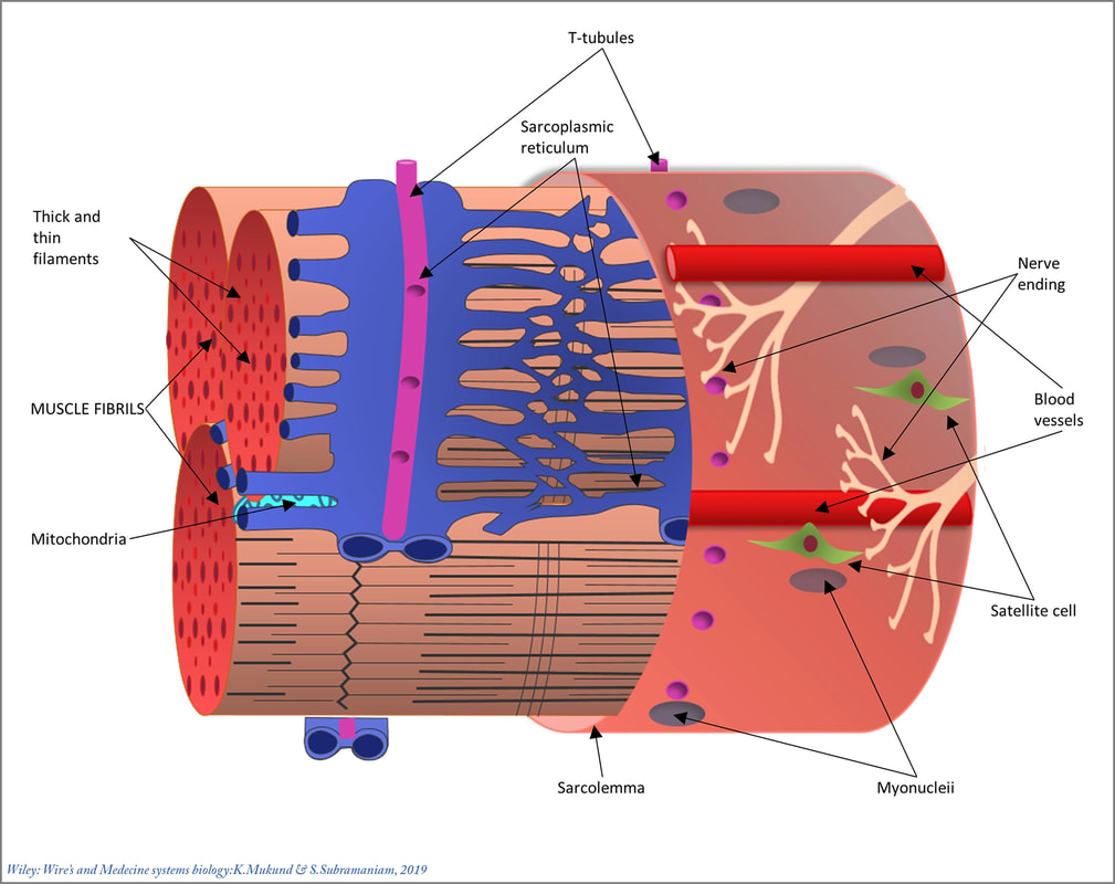
Title: Scheme of the structure of a skeletal muscle fibre
Description:
Each muscle fibre is surrounded by a connective sheath, the endomysium.
The fibres are grouped into bundles (fasciculi) surrounded by the extending connective tissue sheaths, called perimysium.
A muscle as a whole comprises several fasciculi and is surrounded by the connective tissue sheath called epimysium.
Each muscle is supplied by motor and/or sensory nerve fibres entering the deep surface of the muscle and ending in a synaptic contact on the muscle cell (motor unit). Denervated muscle fibres gradually develop atrophy. The force of a muscle contraction is built up by both increasing the frequency of activation of a single motor nerve fibre and by adding more activated nerve fibres simultaneously.
The muscle fibres are also supplied by a rich vascular network.
The contraction of muscle fibres is carried out by myofilaments, consisting of thick and thin filaments organized in the subcellular sarcomere unit, being a construction of contractile filaments such as actin, myosin and many other associated proteins, described in detail elsewhere in this and other sections. The myofilaments are surrounded by mitochondria and glycogen granules for the energy production needed for contraction. The cell membrane or sarcolemma envelops the muscle fibre and penetrates with T-tubules (transverse tubules) deep between the myofibrils in order to propagate the axon innervation trigger into the cell.
In the sarcoplasm, the cisterns of an extended sarcoplasmic reticulum system are assembled near and around the transverse tubules, thus facilitating the Ca++ ions storage and release during the running action potential.
Keywords/Mesh: locomotor system, skeletal muscle, striated muscle, epimysium, perimysium, endomysium, contraction, sarcomere, sarcoplasmic reticulum, transverse tubule, T-tubule, electron microscopy, histology, POJA collection
From: (Skeletal muscle: A review of molecular structure and function, in health and disease, Kavitha Mukund and Shankar Subramaniam, First published: 13 August 2019. https://doi.org/10.1002/wsbm.1462 (open access article). © 2019 The Authors. WIREs Systems Biology and Medicine published by Wiley Periodicals, Inc. This is an open access article under the terms of the Creative Commons Attribution License, which permits use, distribution and reproduction in any medium, provided the original work is properly cited.
Title: Scheme of the structure of a skeletal muscle fibre
Description:
Each muscle fibre is surrounded by a connective sheath, the endomysium.
The fibres are grouped into bundles (fasciculi) surrounded by the extending connective tissue sheaths, called perimysium.
A muscle as a whole comprises several fasciculi and is surrounded by the connective tissue sheath called epimysium.
Each muscle is supplied by motor and/or sensory nerve fibres entering the deep surface of the muscle and ending in a synaptic contact on the muscle cell (motor unit). Denervated muscle fibres gradually develop atrophy. The force of a muscle contraction is built up by both increasing the frequency of activation of a single motor nerve fibre and by adding more activated nerve fibres simultaneously.
The muscle fibres are also supplied by a rich vascular network.
The contraction of muscle fibres is carried out by myofilaments, consisting of thick and thin filaments organized in the subcellular sarcomere unit, being a construction of contractile filaments such as actin, myosin and many other associated proteins, described in detail elsewhere in this and other sections. The myofilaments are surrounded by mitochondria and glycogen granules for the energy production needed for contraction. The cell membrane or sarcolemma envelops the muscle fibre and penetrates with T-tubules (transverse tubules) deep between the myofibrils in order to propagate the axon innervation trigger into the cell.
In the sarcoplasm, the cisterns of an extended sarcoplasmic reticulum system are assembled near and around the transverse tubules, thus facilitating the Ca++ ions storage and release during the running action potential.
Keywords/Mesh: locomotor system, skeletal muscle, striated muscle, epimysium, perimysium, endomysium, contraction, sarcomere, sarcoplasmic reticulum, transverse tubule, T-tubule, electron microscopy, histology, POJA collection