
15.1 POJA-L7005+7004 Hyaline cartilage
15.1 POJA-L7005+7004 Hyaline cartilage
15.1 POJA-L7005+7004 Hyaline cartilage
Title: Hyaline cartilage
Description:
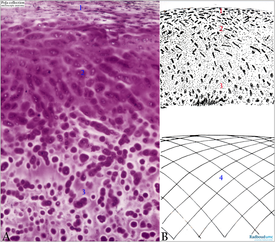
(A): Basic fuchsin staining of human hyaline articular cartilage.
(1): Perichondrium with small spindle-like chondroblasts that grow and differentiate into chondrocytes (2) embedded in dark pink stained matrix of proteoglycans and glycoproteins (glycosaminoglycans, chondroitin sulphates, keratan sulphates and hyaluronic acids). This type of growth derived from the perichondrium is the appositional growth in contrast to the interstitial growth that occurs within the isogenic groups of chondrocytes. The growth is very limited in adults. Collagen fibres are masked within the matrix. In the deeper part (3) the staining is less intense and the fibrillar aspect of the matrix is partly exposed.
(B): Scheme of cartilage at the top:
(1): Superficial zone;
(2): Middle zone;
(3): Deep zone, illustrating the formation of isogenic groups of cartilage cells (2) derive from the perichondrial chondroblasts (1).
Close to (3) at the bottom part of fibrillated matrix.
(4): Scheme at the bottom propose the collagenous arcadic architecture responsible for the contribution to tensile strength of this type of cartilage. Area 4 is comparable to that of the area between 2 and 3 in the top scheme.
Background:
Chondrocytes and their pericellular matrix in cartilage are organized as chondrons which consist of the chondrocyte and the pericellular molecular proteins of which collagen type VI and IX are the major components. Chondrons are the primary structural, functional, and metabolic units in a cartilage. In a chondron the chondrocyte maintains a stable genotype.
The biomechanical properties of cartilage in joints are both poro-viscoelastic (fluid flow dependent mechanism) and depth-dependent (fluid flow independent) as a result of the unique molecular and morphological structures of articular cartilage.
References:
Keywords/Mesh: locomotor system, cartilage, hyaline, matrix, collagen fibre, histology, POJA collection
Title: Hyaline cartilage
Description:
(A): Basic fuchsin staining of human hyaline articular cartilage.
(1): Perichondrium with small spindle-like chondroblasts that grow and differentiate into chondrocytes (2) embedded in dark pink stained matrix of proteoglycans and glycoproteins (glycosaminoglycans, chondroitin sulphates, keratan sulphates and hyaluronic acids). This type of growth derived from the perichondrium is the appositional growth in contrast to the interstitial growth that occurs within the isogenic groups of chondrocytes. The growth is very limited in adults. Collagen fibres are masked within the matrix. In the deeper part (3) the staining is less intense and the fibrillar aspect of the matrix is partly exposed.
(B): Scheme of cartilage at the top:
(1): Superficial zone;
(2): Middle zone;
(3): Deep zone, illustrating the formation of isogenic groups of cartilage cells (2) derive from the perichondrial chondroblasts (1).
Close to (3) at the bottom part of fibrillated matrix.
(4): Scheme at the bottom propose the collagenous arcadic architecture responsible for the contribution to tensile strength of this type of cartilage. Area 4 is comparable to that of the area between 2 and 3 in the top scheme.
Background:
Chondrocytes and their pericellular matrix in cartilage are organized as chondrons which consist of the chondrocyte and the pericellular molecular proteins of which collagen type VI and IX are the major components. Chondrons are the primary structural, functional, and metabolic units in a cartilage. In a chondron the chondrocyte maintains a stable genotype.
The biomechanical properties of cartilage in joints are both poro-viscoelastic (fluid flow dependent mechanism) and depth-dependent (fluid flow independent) as a result of the unique molecular and morphological structures of articular cartilage.
References:
- Yang Xia, Konstantin I. Momot, Zhe Chen, Christopher T. Chen, David Kahn and Farid Badar, CHAPTER 1:Introduction to Cartilage in Biophysics and Biochemistry of Cartilage by NMR and MRI, 2016, pp. 1-43 DOI: 10.1039/9781782623663-00001
- The poro-viscoelastic properties of trabecular bone: a micro computed tomography-based finite element study. Clara Sandino 1 , David D McErlain 1 , John Schipilow 2 , Steven K Boyd 3 J Mech Behav Biomed Mater . 2015 Apr;44:1-9. https://doi.org/10.1016/j.jmbbm.2014.12.018
Keywords/Mesh: locomotor system, cartilage, hyaline, matrix, collagen fibre, histology, POJA collection