
8.2 POJA-L309
Survey of the nasal conchae (isolated turbinate bones)
8.2 POJA-L316.
Nasal concha (isolated turbinate bone)
8.2 POJA-L317.
Nasal concha with respiratory mucosa
|
8.2 POJA-L309
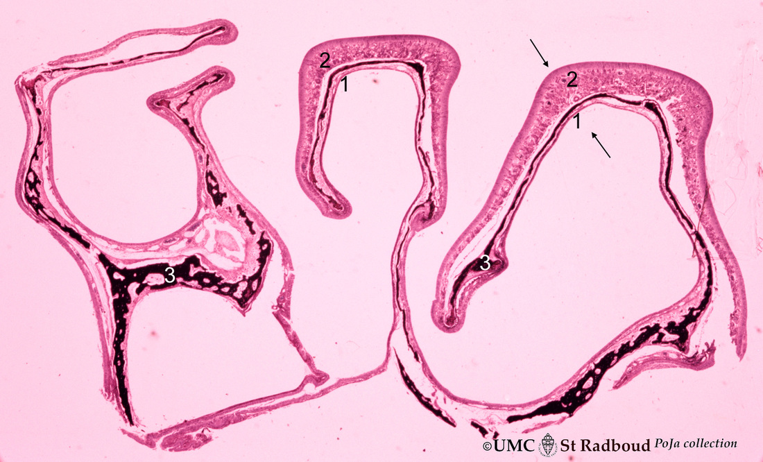
Title: Survey of the nasal conchae (isolated turbinate bones) Description: Satin hematoxylin-eosin, dog. The conchae (turbinates) consist of three parts: the inferior, middle and superior turbinate bones (3, black-stained); they are covered by a respiratory mucosa (1) or by an olfactory mucosa (2). In human only a small part of the superior concha exhibits olfactory epithelium in contrast to the dog where large olfactory areas are depicted. Note the differences in thickness between the olfactory epithelium and the respiratory epithelium as indicated by arrows. 8.2 POJA-L316
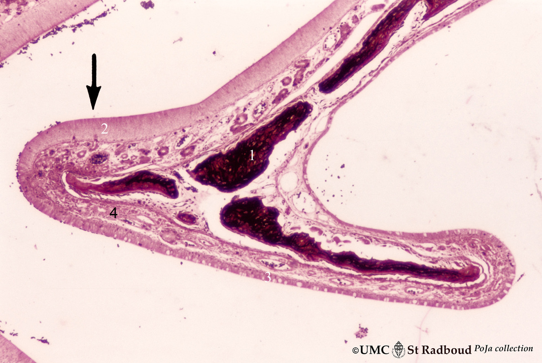
Title: Nasal concha (isolated turbinate bone) Description: Stain hematoxylin-eosin, dog. This concha (turbinate with red-black-stained bone, 1) is covered by a thin respiratory mucosa (3) and by a thick olfactory mucosa (2, arrow). Within the respiratory epithelium light stained goblet cells are visible between the low columnar/cuboidal epithelial cells (cilia not visible at this magnification). The olfactory epithelium consists of tall columnar cells. Blood vessels and small glands are present in the submucosa (4). 8.2 POJA-L317
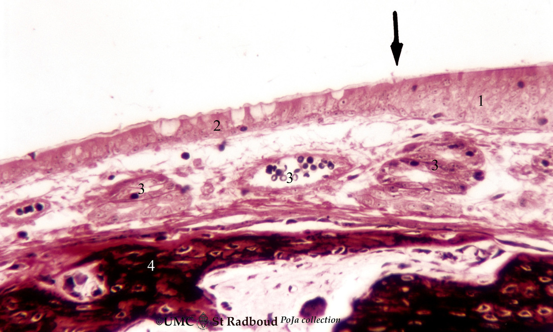
Title: Nasal concha with respiratory mucosa Description: Stain hematoxylin-eosin, dog () Indicates the transition of the olfactory epithelium (1) into the respiratory epithelium (2) with pseudostratified ciliated epithelium and goblet cells. The submucosa is richly vascularized (3). Lamellar bone of the turbinate (4) is stained reddish-black. |
8.2 POJA-L319.
Respiratory epithelial cells in the nasal concha
|
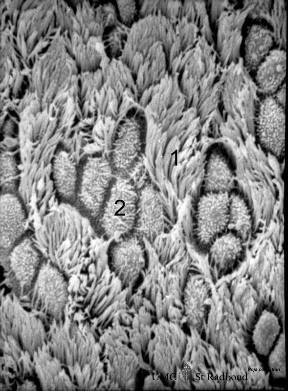
8.2 POJA-L319
Title: Respiratory epithelial cells in the nasal concha Description: Scanning electron microscopy, rat. Bushes of long cilia (1) are present on the top of the epithelial cells. In between clusters of microvilli-studded goblet cells (2) are visible. KeywordsMesh: respiratory tract, nasal cavity, concha, turbinate bone, olfactory epithelium, respiratory epithelium, goblet cell, cilium, microvillus, histology, electron microscopy, POJA collection |