14.1 POJA-L6330+6331+6332+6333 Histochemistry-II of skeletal muscles (human)
14.1 POJA-L6330+6331+6332+6333 Histochemistry-II of skeletal muscles (human)
(By courtesy of H. ter Laak PhD Section Neuropathology, retired staff member Department of Pathology, Radboud university medical center, Nijmegen, The Netherlands)
Title: Histochemistry-II of skeletal muscles (human)
Description:
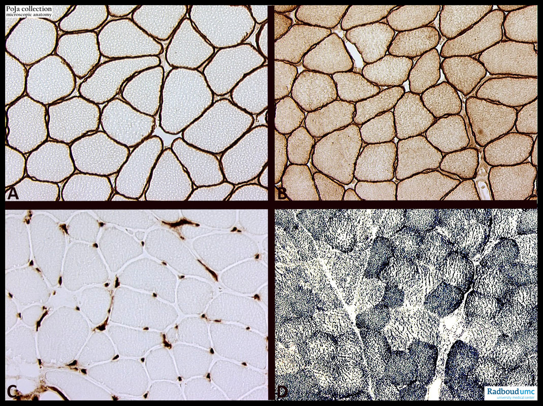
(A): α -Sarcoglycan immunoperoxidase staining.
(B): Caveolin immunoperoxidase staining.
(C): HLA-DR immunoperoxidase staining.
(D): SDH, formazan staining.
The sarcoglycans are a family of single pass transmembrane proteins that are part of the dystrophin-associated glycoprotein complex that links the actin cytoskeleton to the extracellular matrix in cardiac and skeletal muscle, conferring structural stability to the sarcolemma and protecting muscle fibres from mechanical stress during muscle contraction.
The absence of α -sarcoglycan can be detected by immunohistochemistry in cases of limb-girdle dystrophies. These disorders with either autosomal dominant or autosomal recessive inheritance are characterized by a primary protein defect such as e.g. sarcoglycan.
In the skeletal muscle tissue, caveolin-3 is localised along the sarcolemma except for the transverse tubules, and co-immunolocalised with dystrophin, whereas caveolin-1 is absent except in the blood vessels of the muscle tissue. In cardiac muscle cells, caveolins-1 and -3 and dystrophin were co-immunolocalised on the sarcolemma and transverse tubules. In uterine smooth muscle cells, caveolin-1, but not caveolin-3, was co-immunolocalised with dystrophin on the sarcolemma.
Mutations in the gene encoding caveolin-3 (CAV3) is also responsible for limb-girdle muscular dystrophy (type 1C) and cardiomyopathies. Caveolin-3 is involved in membrane repair, when CAV3 is mutated the plasma membrane shows ultrastructural abnormalities.
The myofibres in muscle biopsy specimens with a pronounced inflammatory component can express class II histocompatibility antigens (HLA-DR) by immunohistochemical techniques. Normal myofibres, however, are usually negative. Skeletal muscle fibres in inflammatory myopathies (e.g., polymyositis) express MHC class II as well as MHC class I. The MHC antigen expression is independent of the inflammatory cell infiltration.
Succinate dehydrogenase (SDH) is a mitochondrial enzyme complex located in the inner mitochondrial membrane that has dual roles in the oxidation of succinate to fumarate in the Krebs cycle and in electron transport during oxidative phosphorylation. SDH histochemistry with formazan highlights the oxidative capacity of muscle fibres and therefore allows for the differentiation of type I, or high oxidative capacity (dark staining), and type II, or low oxidative capacity (light staining), fibres. Additionally, abnormal subsarcolemmal mitochondrial aggregates are readily visualized on SDH histochemical stains in cases of mitochondrial myopathies. These abnormal fibres often have the appearance of “ragged-red fibres”.
See also:
Keywords/Mesh: locomotor system, skeletal muscle, striated muscle, sarcolemma, sarcoglycan, caveolin, mitochondrion, succinate dehydrogenase, enzyme, histochemistry, histology, POJA-collection
Title: Histochemistry-II of skeletal muscles (human)
Description:
(A): α -Sarcoglycan immunoperoxidase staining.
(B): Caveolin immunoperoxidase staining.
(C): HLA-DR immunoperoxidase staining.
(D): SDH, formazan staining.
The sarcoglycans are a family of single pass transmembrane proteins that are part of the dystrophin-associated glycoprotein complex that links the actin cytoskeleton to the extracellular matrix in cardiac and skeletal muscle, conferring structural stability to the sarcolemma and protecting muscle fibres from mechanical stress during muscle contraction.
The absence of α -sarcoglycan can be detected by immunohistochemistry in cases of limb-girdle dystrophies. These disorders with either autosomal dominant or autosomal recessive inheritance are characterized by a primary protein defect such as e.g. sarcoglycan.
In the skeletal muscle tissue, caveolin-3 is localised along the sarcolemma except for the transverse tubules, and co-immunolocalised with dystrophin, whereas caveolin-1 is absent except in the blood vessels of the muscle tissue. In cardiac muscle cells, caveolins-1 and -3 and dystrophin were co-immunolocalised on the sarcolemma and transverse tubules. In uterine smooth muscle cells, caveolin-1, but not caveolin-3, was co-immunolocalised with dystrophin on the sarcolemma.
Mutations in the gene encoding caveolin-3 (CAV3) is also responsible for limb-girdle muscular dystrophy (type 1C) and cardiomyopathies. Caveolin-3 is involved in membrane repair, when CAV3 is mutated the plasma membrane shows ultrastructural abnormalities.
The myofibres in muscle biopsy specimens with a pronounced inflammatory component can express class II histocompatibility antigens (HLA-DR) by immunohistochemical techniques. Normal myofibres, however, are usually negative. Skeletal muscle fibres in inflammatory myopathies (e.g., polymyositis) express MHC class II as well as MHC class I. The MHC antigen expression is independent of the inflammatory cell infiltration.
Succinate dehydrogenase (SDH) is a mitochondrial enzyme complex located in the inner mitochondrial membrane that has dual roles in the oxidation of succinate to fumarate in the Krebs cycle and in electron transport during oxidative phosphorylation. SDH histochemistry with formazan highlights the oxidative capacity of muscle fibres and therefore allows for the differentiation of type I, or high oxidative capacity (dark staining), and type II, or low oxidative capacity (light staining), fibres. Additionally, abnormal subsarcolemmal mitochondrial aggregates are readily visualized on SDH histochemical stains in cases of mitochondrial myopathies. These abnormal fibres often have the appearance of “ragged-red fibres”.
See also:
- 14.6.1 POJA-L6190B+6188+6189 Mitochondrial myopathy
- 14.1 POJA-L6326+6327+6328+6329 Histochemistry-I of skeletal muscles
Keywords/Mesh: locomotor system, skeletal muscle, striated muscle, sarcolemma, sarcoglycan, caveolin, mitochondrion, succinate dehydrogenase, enzyme, histochemistry, histology, POJA-collection

