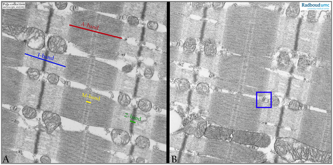
14.1 POJA-L6236+6237 Electron micrograph of sarcomeres in skeletal muscle (human)
14.1 POJA-L6236+6237 Electron micrograph of sarcomeres in skeletal muscle (human)
Title: Electron micrograph of sarcomeres in skeletal muscle (human)
Description:
The electron micrographs of skeletal muscle fibres illustrate the structure of sarcomere units.
The I-band: actin myofilaments (blue bar in A)
The A-band: myosin + actin filaments
The M-band: Myosin filaments in register held by M-line proteins (myomesin, obscurin and M-protein) (yellow in A)
The Triad: complex of T -tubule and two adjacent terminal cisternae at the level of junction between A-band and I-band (A-I junction) in human striated muscle (blue square)
In between the bundles of myofilaments mitochondria of variable sizes and aspects.
Keywords/Mesh: locomotor system, skeletal muscle, striated muscle, sarcomere, A-band, I-band, H-band, M-band, Z-line, triad, electron microscopy, POJA collection

