4.1.1 POJA-L3954+3961+La0138+3953
Title: Cardia part of the esophagus and stomach area
Description:
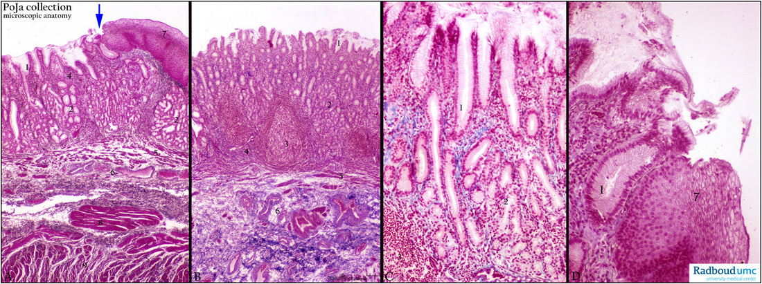
Stain Azan, human. (A, B) Survey. (C, D) Detail.
The esophageal tube is covered inside with a non-keratinizing multilayered squamous epithelium (A, 7 and D, 7) that abruptly converts into a simple cuboidal-columnar epithelium (arrow, ↘) in the stomach (B, 1) and (D). In the stomach they form crypts or foveolae (1) in which end the esophageal glands (2) that produce mucus (and lysozyme) facilitating the passage of food.
The glandular tubules are often convoluted (A) and branched (B).
Note that the cardia glands in esophagus and stomach are continuous and identical.
The loose connective tissue of the lamina propria (4) can locally be filled up with lymph follicles (3) for the immune defense. (5) Lamina muscularis mucosae. Many blood vessels, nerve fibers and some ganglion cells are spread in the tela submucosa (6).
Keywords/Mesh: esophagus, cardia, stomach, glands, lymphoid follicles, histology, POJA collection

