13.1 POJA-La0299+L4579+4581+4580
Title: Ultrastructure of the intima of muscular artery
Description:
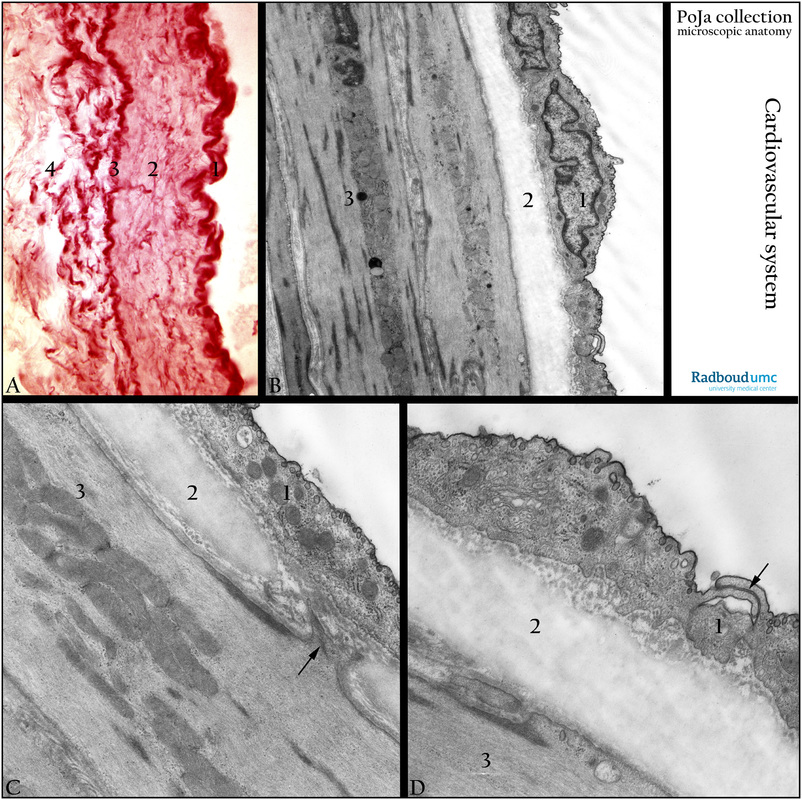
(A): Orcein staining of a small/mid-sized muscular artery, human. (1) Tunica intima with endothelium and internal elastic lamina (IEL). (2) Tunica media with smooth muscle cells and orcein stained dense-red fine elastic fibers. (3) External elastic lamina, followed by adjacent tunica adventitia (4) with coarse elastic fibers.
(B): Electron micrograph mid-sized muscular artery, gold hamster. (1) Endothelial cell resting on a basal lamina followed by an internal elastic lamina (IEL, 2), all elastic laminae are fenestrated. The tunica media (3) mainly contains smooth muscle cells equipped with characteristic dense plaques and filaments. (See C and D).
(C, D): Electron micrograph muscular artery with IEL, endothelial-myocyte junction, golden hamster. (1, 2, 3) similar to (C). Arrow is an endothelial- myocyte junction. The endothelial cells possess a Golgi area, caveolae and pinocytotic vesicles and a thin basal lamina.
Arrow in (D) is an endo-endothelial junction.
Keywords/Mesh: cardiovascular system, vascularisation, blood vessel, muscular artery, intima, elastic lamina, smooth muscle, dense plaque, histology, electron micrograph, POJA collection

