
11.4 POJA-L4444+3088+
3089+4446.
Cerebellum 2
11.4 POJA-L4444+3088+3089+4446
Title: Cerebellum 2
Description:
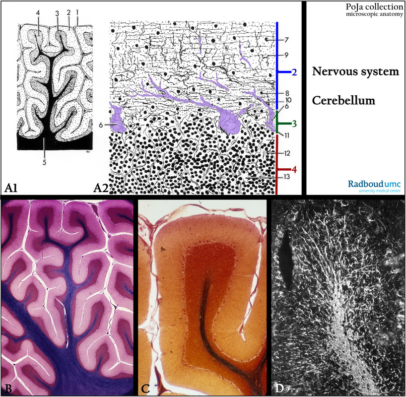
(A1, A2): Scheme structure cerebellum, human. The cortex is the most superficial part is occupied by gray matter,
the central region of the area is the medulla, contains white matter made up of fiber tracts and clusters of nerve cells.
See also Section 11.1
(1) Pia mater.
(2) Stratum moleculare or molecular layer.
(3) Stratum gangliosum (ganglionare) between granular and molecular layer, contains the Purkinje cells (painted pink).
(4) Stratum granulosum or granular layer.
(5) Medulla.
(6) Purkinje cells arranged in a row in the stratum ganglionare. An elaborate branch of dendrites (tree-like) extents deeply in the molecular layer.
(7) Basket cells.
(8) Nucleus glia cell.
(9) Parallel fibers (axons of granule cells in 12).
(10) Dendrites derived from the Purkinje cells with climbing fibers.
(11) Basket cells (with tangential fibers) surrounding the cell body of the Purkinje cells.
(12) Granule cells.
(13) Cerebellar “glomerulus” or parenchym island being the contact place between moss fibers and granule cells.
(B): Modified Weigert staining revealing the so called “Arbor Vitae”, human. The cerebellar cortex is deeply grooved and appears as thin, transverse leaves called folia. Equivalent to (A1).
(C): Silver stain, human. Equivalent to the scheme in (A2).
Note that rests of the arachnoid and pia mater are present.
(D): Immunofluorescent staining with antibodies against neurofilaments , black-white print, mouse. Showing that the filaments are part of the white matter (medulla) and extent deeply into the molecular layer.
Keywords/Mesh: nervous tissue, cerebellum, molecular layer, Purkinje cell layer, granular layer, neurofilament, histology, POJA collection
Title: Cerebellum 2
Description:
(A1, A2): Scheme structure cerebellum, human. The cortex is the most superficial part is occupied by gray matter,
the central region of the area is the medulla, contains white matter made up of fiber tracts and clusters of nerve cells.
See also Section 11.1
(1) Pia mater.
(2) Stratum moleculare or molecular layer.
(3) Stratum gangliosum (ganglionare) between granular and molecular layer, contains the Purkinje cells (painted pink).
(4) Stratum granulosum or granular layer.
(5) Medulla.
(6) Purkinje cells arranged in a row in the stratum ganglionare. An elaborate branch of dendrites (tree-like) extents deeply in the molecular layer.
(7) Basket cells.
(8) Nucleus glia cell.
(9) Parallel fibers (axons of granule cells in 12).
(10) Dendrites derived from the Purkinje cells with climbing fibers.
(11) Basket cells (with tangential fibers) surrounding the cell body of the Purkinje cells.
(12) Granule cells.
(13) Cerebellar “glomerulus” or parenchym island being the contact place between moss fibers and granule cells.
(B): Modified Weigert staining revealing the so called “Arbor Vitae”, human. The cerebellar cortex is deeply grooved and appears as thin, transverse leaves called folia. Equivalent to (A1).
(C): Silver stain, human. Equivalent to the scheme in (A2).
Note that rests of the arachnoid and pia mater are present.
(D): Immunofluorescent staining with antibodies against neurofilaments , black-white print, mouse. Showing that the filaments are part of the white matter (medulla) and extent deeply into the molecular layer.
Keywords/Mesh: nervous tissue, cerebellum, molecular layer, Purkinje cell layer, granular layer, neurofilament, histology, POJA collection