
2.1 POJA-L901. Thymus (human, newborn)
2.1 POJA-L901
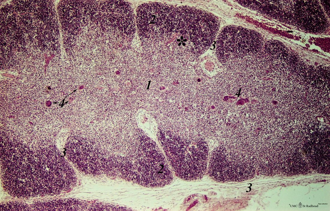
Title: Thymus (human, newborn)
Description: Stain: Hematoxylin & eosin.
The thymus is a bilobed lymphoepithelial organ derived as an outgrowth from the third branchial (pharyngeal) pouch, and situated in the anterior mediastinum.
Each lobe is divided into multiple lobules by fibrous septa or trabeculae (3). Each lobule consists of an outer cortex (2) and an inner medulla (1).
The cortex contains a dense collection of immature T lymphocytes, and the lighter staining medulla is more sparsely populated with mature, immunocompetent T cells. Only mature T cells exit the thymus and enter the blood and peripheral lymphoid organs.
Scattered throughout the thymus are non-lymphoid epithelial cells, as well as bone marrow-derived macrophages and dendritic cell types. These dendritic cells contribute to the proliferation, maturation and selection process of the thymocytes.
In the medulla are structures called Hassall’s corpuscles (4), which are composed of tightly packed whorls of epithelial reticular cells that may be remnants of degenerating, keratinized cells. Thymic hormones or growth factors are found in the Hassall bodies, such as thymosin and thymopoietin, thymulin. The thymus has a rich vascular supply (3) and efferent lymphatic vessels that drain into the mediastinal lymph nodes. A fine capillary network exists at the transition zone (*) between cortex and medulla. These capillaries are ensheathed by basal laminae and branched epithelial reticular cells forming part of the so-called blood-thymus barrier.
Keywords/Mesh: lymphatic tissue, thymus, Hassall’s corpuscle, thymic corpuscle, histology, POJA collection