4.1.1 POJA-L3947+3948+3949
Title: Plexus of Auerbach in digestive tract (human, rat)
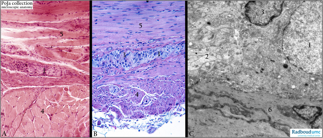
Description: (A) Hematoxylin-eosin, human. (B) Hematoxylin-eosin-Weigert. (C) Electron micrograph ganglion cell (1), interstitial cell (3) (former mantle cells) in jejunum, rat.
The plexus of Auerbach or myenteric plexus (1) is located between the two muscle layers (4, 5) of the tunica muscularis (circular and longitudinal), in the whole digestive tract. The plexus consists of ganglion cells (C, 1) surrounded by interstitial cells (former mantle layer cells) (C, 3) and nerve branches as shown in (C, 2). Smooth muscle cells with dark small dense plaques (C, 6). The dense plaques (containing vinculin, talin and the fibronectin receptor) are anchorage sites of the contractile apparatus to the membrane and matrix.
Keywords/Mesh: plexus of Auerbach, ganglion cells, digestive tract, electron microscopy, histology, POJA collection

