14.3 POJA-L6018+6019 Myoepithelial cells in sweat glands (human)
14.3 POJA-L6018+6019 Myoepithelial cells in sweat glands (human)
Title: Myoepithelial cells in sweat glands (human)
Description:
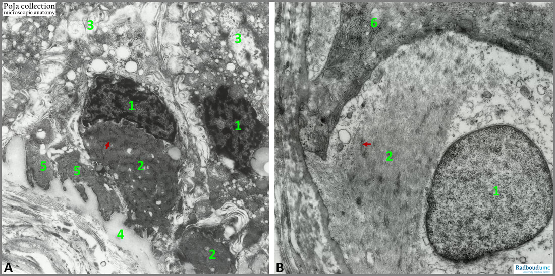
(A): Survey basal part of a sweat gland. (1): Nuclei of glycogen-rich cells (so-called “clear cells” in light microscopy). (2): Its cytoplasm with glycogen and dark mitochondria. (3): Widened intercellular spaces between “clear cells”. Basally close to the lamina propria (4) dark remnants (5) of myoepithelial cells resting on a basal lamina (zoom in!).
(B): Detail of myoepithelial cell. (1): Nucleus of myoepithelial cell with myofilaments (actin) (2) and dense plaques or bodies (arrows).
(6): Part of a gland cell.
Functionally myoepithelial cell contractions are similar to those of smooth muscle cells and are capable to squeeze out the secretion products of the active gland cells.
In the body there are various contractile cells that contain distinct amount of actin. In their functional activity they resemble smooth muscle cells. Examples are:
1. Vascular pericytes described in
In wounds or bone fractures so-called free myofibroblasts proliferate during the healing processes.
Contractile cells can display also epithelial characteristics such as myoepithelial cells found in:
4. Mammary glands
Keywords/Mesh: locomotor system, sweat gland, smooth muscle cell, myoepithelial cell, electron microscopy, histology, POJA collection
Description:
(A): Survey basal part of a sweat gland. (1): Nuclei of glycogen-rich cells (so-called “clear cells” in light microscopy). (2): Its cytoplasm with glycogen and dark mitochondria. (3): Widened intercellular spaces between “clear cells”. Basally close to the lamina propria (4) dark remnants (5) of myoepithelial cells resting on a basal lamina (zoom in!).
(B): Detail of myoepithelial cell. (1): Nucleus of myoepithelial cell with myofilaments (actin) (2) and dense plaques or bodies (arrows).
(6): Part of a gland cell.
Functionally myoepithelial cell contractions are similar to those of smooth muscle cells and are capable to squeeze out the secretion products of the active gland cells.
In the body there are various contractile cells that contain distinct amount of actin. In their functional activity they resemble smooth muscle cells. Examples are:
1. Vascular pericytes described in
- 13.1 POJA-L4642+La0301+4721+4626+4625 Capillaries: inner and outer lining cells
- 13.1 POJA-L4592+4600+4632+4601+4635+4636+La0297 Arterioles
- 6.1 POJA-L2671+2672+2680 Blood-testis barrier
- 11.2.2 POJA-L2076+3280+3281+4310 +4311+3282+3283 Sensory receptors (Vater-Pacini/Meissner’s corpuscles
- 11.2 POJA-L3256+3273+3229+3453 Myelinated peripheral nerve fibers 7
- 10.3 POJA-L2037+2242+2080+2087+2243+2089 Vater-Pacini corpuscles I in the skin
- 5.7 POJA-L5024+5025+5026+5027 Urinary bladder wall and lining in sheep
In wounds or bone fractures so-called free myofibroblasts proliferate during the healing processes.
Contractile cells can display also epithelial characteristics such as myoepithelial cells found in:
4. Mammary glands
- 7.4.1 POJA-L1643+1943+103 Mammary gland in pregnancy
- 10.4 POJA-L2085+2086+2201+2269+2186+2205+2207 Eccrine sweat glands and myoepithelial cells
Keywords/Mesh: locomotor system, sweat gland, smooth muscle cell, myoepithelial cell, electron microscopy, histology, POJA collection

