
12.2.4.1 POJA-L4340+3419+
3422+3415+2986
Development of the organ of Corti in the inner ear
12.2.4.1 POJA-L4340+3419+3422+3415+2986
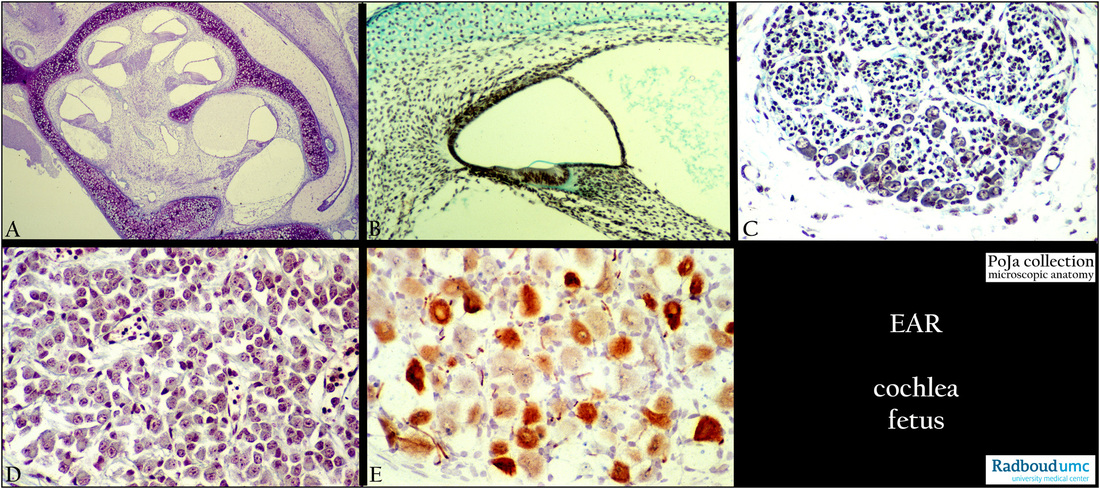
Title: Development of the organ of Corti in the inner ear
Description:
(A): Survey cochlea, stain toluidine blue, 4 days postnatal rat.
(B): Ductus cochlearis, stain trichrome Goldner, fetus human. Immature organ of Corti, note the well-defined thin blue-green tectorial
membrane produced by the interdental cells.
(C): Spiral ganglion with nerve fibres and peripheral ganglion cells, stain trichrome Goldner, fetus human.
(D): Spiral ganglion, stain trichrome Goldner, fetus human.
(E): Spiral ganglion, immunoperoxidase staining with AEC and antibodies against neurofilament, 4 days postnatal rat.
There is varying intensity of reactivity of the ganglion cells during development.
Keywords/Mesh: fetus, inner ear, cochlea, cochlear duct, spiral ganglion, neurofilament, histology, POJA collection