
2.3 POJA-L1013. Lymph node (human)
2.3 POJA-L1013
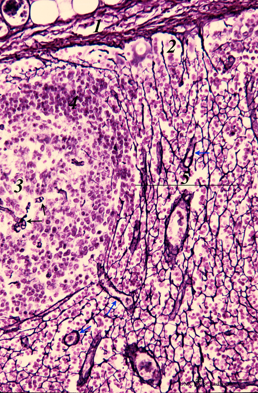
Title: Lymph node (human)
Description: Stain: Gomori silver. Due to the argyrophilia of reticular fibres the reticular network is black-stained among other fibers such as collagen type III, IV.
From the fibrous capsule (1) a meshwork of fine reticular fibres penetrates into the cortex of a lymph node crossing the subcapsular (or marginal) sinus (2). At the left a secondary lymphatic nodule (or follicle) is shown with centrally a germinal centre (3) with capillaries (→). This (secondary) gc is surrounded by a dense cap or mantle zone (4) of B-lymphocytes. Note that the reticular fibers are nearly absent in the germinal centre.
(5) Area of intermediate sinus (or paratrabecular sinuses) stuffed with lymphocytes.
(Blue arrows) indicate small arterioles with black-stained basement membranes (collagen IV).
Keywords/Mesh: lymphatic tissue, lymph node, reticular tissue, argyrophilia, lymphatic follicle, histology, POJA collection