10.7 POJA-L4347+3886+4350+3887+4351
Title: Seborrheic keratosis (verruca seborrhoica) I
Description:
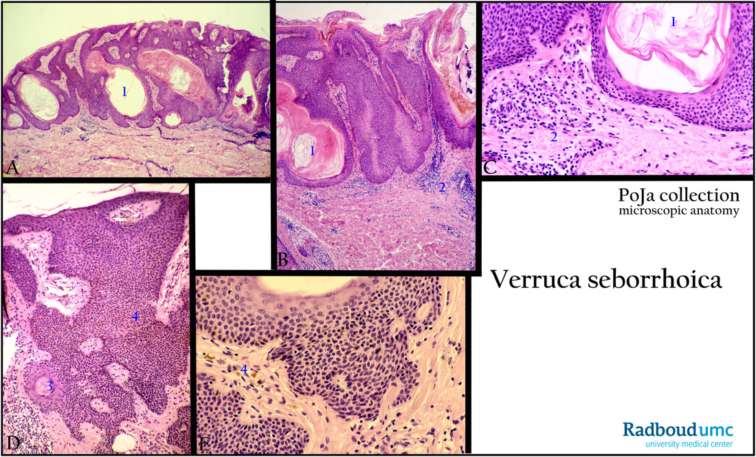
(A): Exophytic growth pattern of this benign neoplasma, the epithelium raises above the rest of the skin, stain hematoxylin-eosin, human.
Note pseudo-horn cysts (1).
(B): Hyperkeratosis on the surface and large pseudo-horn cysts as a result of down growths of keratin into the main tumor mass.
The epithelium is papillomatous and hyperkeratotic.
(C): Small basophilic tumor cells and keratin. (2) Infiltrate.
(D, E): The tumor is composed of sheets of cells resembling the basal cells of the normal epidermis. (3) Small horn cyst.
(4) Varying melanin pigmentation contributes to the brown colour (zoom in!!).
Small basophilic tumor cells resembling basal cells of the epidermis.
(Micrographs (D, E) by courtesy of D. Ruiter, MD PhD, former Head Department of Pathology, Radboud university medical center, Nijmegen, The Netherlands)
Background: Spontaneous arising warty growth – most often in middle-aged or elderly – and frequently localized on trunk, face,
back and chest. The tumor manifests itself as flat coin-like plaques uniformly tan or as granular surface lesions up to few centimeters. Sometimes there is clinically confusion with malignant melanoma with the result of excision either for that reason or for cosmetic ones. The tumor rarely progresses as malignant.
Keywords/Mesh: skin, seborrheic keratosis, verruca seborrhoica, basal cell papilloma, histology, pathology, POJA collection

